
Diagnostic Musculoskeletal Ultrasound: Bedside Assessment of Rotator Cuff Tears
In short
High-resolution ultrasound provides real-time dynamic imaging of rotator cuff tears right at the clinical bedside. Discover how clinicians assess tendon integrity.
In this guide (4 sections)
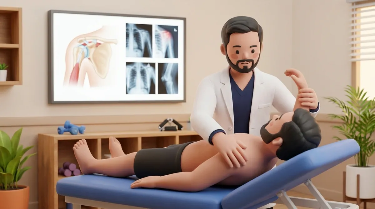
Diagnostic Musculoskeletal Ultrasound (MSK US) has revolutionized the clinical evaluation of rotator cuff pathology, transitioning diagnostic imaging from distant radiology suites directly to the clinical bedside. Utilizing high-frequency linear transducers (twelve to eighteen megahertz), point-of-care ultrasound offers image resolution that rivals or exceeds magnetic resonance imaging for superficial soft tissues. Unlike static MRI scans where the patient lies immobilized inside a magnetic bore, ultrasound permits real-time dynamic examination. The clinician can directly visualize tendon gliding beneath the acromion during active arm movement, assess muscle compressibility, perform real-time stress testing, and compare findings instantaneously with the contralateral asymptomatic shoulder.
Accessible Bedside Imaging in Indian Outpatient Clinical Practice
In India, obtaining an MRI scan often involves significant financial expenditure, long waiting times in corporate diagnostic centres, and travel across congested urban corridors. For elderly individuals or claustrophobic patients, lying motionless inside a noisy scanner drum for forty-five minutes is a distressing experience. Bedside ultrasound offers a non-invasive, cost-effective, and interactive alternative. In urban clinics across Delhi, Mumbai, Bengaluru, and Chennai, physiotherapists and sports physicians utilize point-of-care ultrasound during the initial consultation. The patient sits comfortably upright and observes the live monitor screen while the clinician demonstrates why certain reaching movements, such as lifting water jugs or reaching into low cupboards, trigger localized tendon pinching.
Sonographic Criteria: Hypoechogenicity, Cortical Irregularity, and Dynamic Impingement
Accurate sonographic diagnosis of rotator cuff tears relies on standardized acoustic criteria. A healthy tendon appears as a continuous, tightly packed fibrillar pattern of bright, hyperechoic parallel lines. In partial or full-thickness tears, the structural discontinuity appears as an anechoic (black fluid-filled) or hypoechoic defect that disrupts the normal fibrillar architecture. Secondary sonographic signs provide high diagnostic certainty: focal herniation of the subdeltoid fat pad into the tendon gap, loss of the normal convex outer tendon contour, and pronounced cortical irregularity or pitting of the greater tuberosity footprint. Dynamic evaluation allows the clinician to observe the supraspinatus tendon gliding beneath the coracoacromial ligament during scaption, capturing subtle transient impingement that static MRI cannot display.
Integrating Sonographic Feedback into Criterion-Based Rehabilitation
Bedside ultrasound provides an objective biological monitoring tool that guides the pace of physical rehabilitation. Following surgical repair, ultrasound can confirm structural continuity and tendon healing onto the greater tuberosity before the clinician advances the patient to active resistance exercises. In non-operative management, ultrasound visualizes the reduction of subacromial bursal effusion and verifies the absence of dynamic mechanical abutment as scapular kinematics improve. Furthermore, sonographic biofeedback can be used therapeutically: patients watch their muscle belly contract in real time, accelerating motor learning and recruitment of dormant rotator cuff fibers.
Real-Time Ultrasound Biofeedback External Rotation Setting
Objective: Utilize visual sonographic biofeedback to confirm isolated infraspinatus motor contraction without deltoid substitution
- 1Sit comfortably upright next to the ultrasound monitor with your arm resting at twenty degrees of abduction.
- 2Watch the monitor screen as your clinician positions the transducer over your posterior infraspinatus muscle belly.
- 3Gently rotate your forearm outward against light manual resistance, observing the muscle belly thicken on screen.
- 4Notice the difference on screen when you hike your shoulder blade versus when you isolate the rotator cuff correctly.
- 5Hold the pure infraspinatus contraction for 5 seconds, relax for 3 seconds, and repeat with focused awareness.
| Diagnostic Feature | High-Resolution Musculoskeletal Ultrasound (POCUS) | Standard 1.5T / 3.0T Magnetic Resonance Imaging (MRI) |
|---|---|---|
| Dynamic Movement Assessment | Real-time dynamic visualization during active arm elevation, rotation, and subacromial gliding maneuvers. | Static examination; patient immobilized in supine neutral position; cannot assess dynamic impingement. |
| Spatial Soft Tissue Resolution | Exceptional spatial resolution (< 0.1 mm) for superficial tendon fibrils using 15-18 MHz transducers. | Excellent overall contrast resolution; superior for deep intra-articular labral and bony marrow pathology. |
| Cost & Accessibility in India | Highly cost-effective (typically ₹1,000 to ₹2,500); immediate point-of-care availability during consultation. | Higher out-of-pocket cost (typically ₹5,000 to ₹10,000); requires separate scheduling at imaging centre. |
| Patient Comfort & Safety | Completely non-claustrophobic; quiet upright seated examination; safe with cardiac pacemakers and metal implants. | High claustrophobia rates; loud acoustic noise; contra-indicated in certain non-MRI-compatible metallic implants. |
When performing ultrasound of the rotator cuff, tilting the sound beam away from a perpendicular angle causes an acoustic artifact called anisotropy, where a healthy tendon falsely appears black and torn. Diagnostic interpretation must always verify that suspected defects persist across multiple imaging planes and under transducer rocking.
Frequently Asked Questions
Can an ultrasound show whether my rotator cuff tear has healed after surgery?
Yes. High-resolution ultrasound is highly accurate for checking whether a surgically repaired tendon remains firmly anchored to the bone. It can visualize the surgical suture anchors, confirm continuous tendon fibers, and detect any fluid gaps or re-tears without exposing you to radiation.
Do I need a doctor referral to see a rotator cuff physiotherapist on BookPhysio.in?
BookPhysio.in does not require a physician prescription. Under the National Commission for Allied and Healthcare Professions (NCAHP) Act 2021, patients across India enjoy direct access to autonomous registered physiotherapists. Through BookPhysio.in, you connect directly with verified clinicians and pay per session with transparent fees, completely free from advance package commitments, commission markups, or platform subscription charges.
Related recovery guides & articles
BookPhysio.in Editorial Team
The BookPhysio.in editorial team writes and maintains this content, checking it against reliable clinical sources. Content follows our medical review policy: general information only, and no page claims a clinical review until a named registered physiotherapist has signed it off.
Read our medical review policy