
Gluteal Tendon Partial-Thickness Tears: Conservative Loading Rules and Rehabilitation
In short
A clinical rehabilitation guide for partial-thickness gluteal tendon tears, detailing conservative load management and progressive remodeling rules.
In this guide (4 sections)
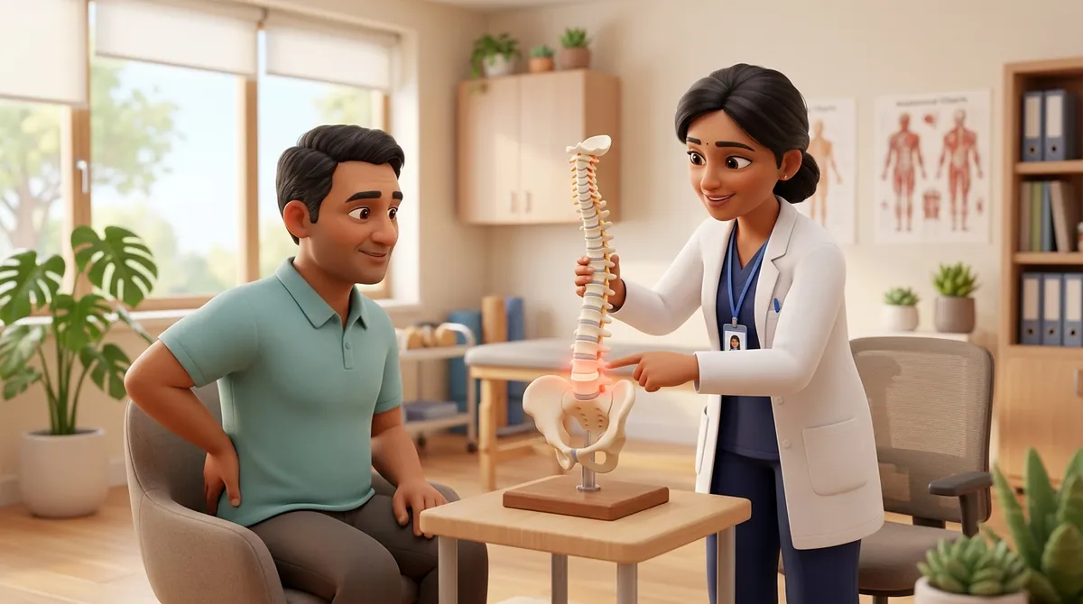
Partial-thickness tears of the gluteus medius and gluteus minimus tendons represent an advanced stage of degenerative tendinopathy, frequently diagnosed on magnetic resonance imaging in middle-aged and older adults presenting with chronic lateral hip pain. These tears typically originate on the deep, articular surface of the tendon footprint, where tissue is subjected to combined tensile and compressive shear over the lateral facet of the greater trochanter. Receiving an MRI report confirming a tendon tear often induces severe fear and kinesiophobia in patients, prompting many to believe that surgery is their only viable option. However, orthopaedic clinical trials consistently show that high-grade partial tears possess substantial conservative healing potential. Because a significant proportion of the tendon's cross-sectional area remains structurally intact, properly dosed mechanotherapy can stimulate remodeling in the surviving fascicles, compensating for the defect and restoring painless, active function without surgical intervention.
Fear of Movement, Squatting Avoidance, and Low Stool Navigation
For an Indian patient diagnosed with a partial gluteal tendon tear, everyday domestic life becomes filled with hesitation and movement apprehension. Traditional daily activities such as squatting to clean floors (jhadu-pocha), getting up from low western or Indian toilets, or lifting heavy stainless steel milk pots are viewed with intense anxiety. Family members, eager to help, often advise the patient to stay in bed and avoid all walking, leading to rapid muscle wasting and cardiovascular deconditioning. When the patient does attempt to move, they walk with a stiff, guarded gait, leaning their upper body heavily over the affected hip to prevent the tendon from working. This protective movement pattern perversely increases compressive adduction moments, worsening lateral pain and confirming the patient's fear that their hip is failing. Overcoming this cycle requires clear clinical education regarding safe loading boundaries.
Intact Fascicle Bridges, Stress Concentration, and 24-Hour Load Latency
The structural pathology of a partial-thickness tear involves a focal discontinuity within the tendon footprint, with longitudinal clefts separating frayed collagen fibers. The primary biomechanical challenge is stress concentration: the tensile load that was previously distributed across the entire broad tendon footprint must now be supported by the remaining intact collagen bridges. If loading is too aggressive or applied at high velocities, these intact bridges can experience tensile yield failure, propagating the tear. Conversely, if the tendon is completely unloaded, the intact fibers undergo disuse atrophy, losing tensile stiffness and matrix cross-links. Successful conservative management hinges on applying controlled mechanical strain that stimulates mechanotransduction in the intact bridges while keeping peak stress safely below their failure threshold. Because tendon tissue exhibits a delayed biological response, symptoms must be monitored over a full twenty-four-hour cycle rather than judging tolerance solely during the exercise itself.
The Safe Loading Traffic Light System, Neutral Range Holds, and Assisted Ambulation
Rehabilitation of partial gluteal tears operates under strict conservative loading rules, often structured as a 'traffic light' system. Green zone loading involves pain between zero and three on a ten-point scale, which is completely safe; amber zone loading involves mild discomfort between four and five that settles within twenty-four hours; red zone loading involves pain exceeding five, or pain that persists into the next morning, indicating tissue overload. All early strengthening exercises must be performed within a neutral joint range, strictly prohibiting any hip adduction or deep flexion that would wrap the torn footprint against bone. During the initial six to eight weeks, patients utilize a single walking stick or crutch in the opposite hand during community ambulation to offload the healing tendon. Isometric holds progress gradually to slow, low-velocity isotonic abductor exercises, rebuilding robust functional capacity over three to six months.
Supported Standing Slider Hip Abduction in Neutral Alignment
Objective: Gently strengthen the intact gluteal tendon fibers in a controlled, non-compressive standing range
- 1Stand tall facing a sturdy table or kitchen counter, resting both hands flat on the surface for balance support.
- 2Place a small plastic paper plate or smooth cloth towel beneath the foot of your painful leg on a slick floor.
- 3Keeping your torso completely upright and your weight resting primarily on your unaffected leg, slowly slide your painful foot outward twelve inches.
- 4Pause for two seconds in this slight abduction position, feeling your outer hip muscles contract smoothly without any pinching.
- 5Slowly slide your foot back to the starting position over three full seconds, ensuring your foot does not cross inward past your midline.
| Monitoring Period | Acceptable Tissue Response (Safe to Progress) | Unacceptable Overload Response (Requires Regression) |
|---|---|---|
| During Exercise Session | Mild muscular effort or dull ache (score 1 to 3/10), no sharp stabbing | Sharp, localized pain directly over the bone (score > 4/10), catching sensations |
| Evening Following Exercise | Comfortable resting in bed, mild muscular fatigue, able to find sleep position | Throbbing pain, unable to lie on either side, requiring pain medications |
| Next Morning Awakening | Morning stiffness resolves within 15 minutes of gentle walking | Marked morning stiffness, severe limping for over 60 minutes, pain on weight-bearing |
| Subsequent 48-Hour Trend | Pain levels return to baseline within 24 hours, functional walking preserved | Elevated baseline pain persisting past 48 hours, loss of single-leg balance capacity |
In patients with documented partial gluteal tears, corticosteroid injections must be avoided. Injecting steroids into or around a structurally compromised tendon severely accelerates collagen necrosis and is one of the leading causes of conversion from a partial-thickness tear into an irreversible full-thickness tendon avulsion. If conservative loading fails over a dedicated six-month period, orthopaedic consultation for trans-osseous surgical repair is indicated.
Frequently Asked Questions
Can a partial tear in my gluteal tendon heal completely without surgery?
While the physical gap of a partial tear may not completely reconnect into pristine original tissue on an MRI, conservative physiotherapy can stimulate the remaining healthy tendon fibers to thicken and remodel. Research demonstrates that the vast majority of patients with partial gluteal tears regain full, pain-free mobility and return to active daily living through progressive strengthening, without ever requiring surgical repair.
Do I need a doctor referral to treat trochanteric hip pain on BookPhysio.in?
BookPhysio.in does not require a doctor referral. Under the National Commission for Allied and Healthcare Professions (NCAHP) Act 2021, patients across India enjoy direct access to autonomous registered physiotherapists. Through BookPhysio.in, you connect directly with verified clinicians and pay per session with transparent fees, completely free from advance package commitments, commission markups, or platform subscription charges.
Related recovery guides & articles
BookPhysio.in Editorial Team
The BookPhysio.in editorial team writes and maintains this content, checking it against reliable clinical sources. Content follows our medical review policy: general information only, and no page claims a clinical review until a named registered physiotherapist has signed it off.
Read our medical review policy