
Heterotopic Ossification Prophylaxis: Gentle Motion vs Forced Stretching
In short
Prevent heterotopic ossification in injured elbows. Understand ectopic bone biology, pharmaceutical prophylaxis, and why forced stretching is banned.
In this guide (4 sections)
Heterotopic ossification (HO) is the pathological formation of mature lamellar bone within extra-skeletal soft tissues, including muscles, tendons, ligaments, and joint capsules. The elbow joint represents the second most frequent site of traumatic heterotopic bone formation in the human body, trailing only the hip. Developing in up to thirty percent of complex elbow fractures, terrible triad injuries, and burn trauma, HO bridges the humeroulnar or proximal radioulnar joints, creating rigid, unbreakable mechanical ankylosis. One of the most catastrophic triggers for ectopic bone formation is aggressive, forceful passive stretching administered by well-meaning but misinformed practitioners. Modern clinical protocols mandate absolute avoidance of forceful manipulation, replacing it with gentle, pain-free active-assisted motion and targeted pharmaceutical prophylaxis.
Unregulated Bone-Setting, Aggressive Massage, and Indian Trauma
In many parts of India, patients recovering from elbow fractures or dislocations frequently consult traditional unregulated bone-setters (pehlwans or jarrahs). These practitioners often subject stiff, post-traumatic elbows to violent, forceful manual manipulation, literally forcing the joint straight against severe patient screams and tears. In other instances, family members vigorously massage the anterior cubital fossa with hot mustard oil or herbal concoctions, applying deep kneading over torn muscle fibers. This traumatic shearing of the brachialis muscle belly causes extensive intramuscular micro-hemorrhage, seeding osteoprogenitor cells and triggering rapid, massive heterotopic bone formation that permanently freezes the elbow.
Ectopic Osteogenesis Biology, BMP Cascades, and Prophylaxis Regimens
The molecular cascade of heterotopic ossification begins with acute local tissue trauma and hypoxemia. Damaged muscle cells and inflammatory macrophages release bone morphogenetic proteins (specifically BMP-2, BMP-4, and BMP-7) and transforming growth factor-beta (TGF-beta). These cytokines stimulate local mesenchymal stem cells located in the perivascular niche of the brachialis muscle to differentiate into osteoblasts, initiating endochondral bone formation within three to six weeks. Pharmaceutical prophylaxis aims to disrupt this inflammatory signaling. Non-steroidal anti-inflammatory drugs (NSAIDs), classically indomethacin (seventy-five milligrams sustained release daily for three to six weeks), inhibit cyclooxygenase (COX-1 and COX-2) enzymes, halting prostaglandin E2 production and blocking osteoblastic differentiation. In high-risk trauma patients, single-fraction low-dose external beam radiation (seven to eight Gray) delivered within seventy-two hours of surgery provides potent local prophylaxis.
Active-Assisted Range of Motion Rules and Cryotherapy Protocols
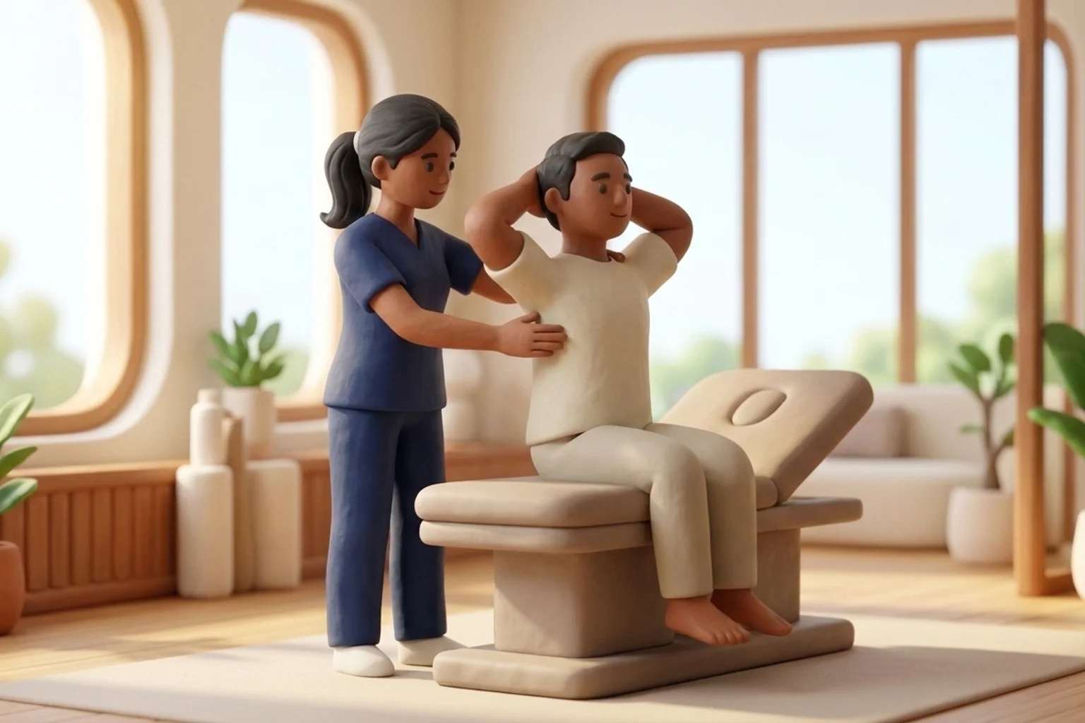
In the rehabilitation clinic, the golden rule of elbow trauma management is absolute gentleness: active and active-assisted motion is encouraged, but passive forced stretching is strictly prohibited. The patient must actively control all range of motion exercises within a completely pain-free arc. If an active exercise provokes sharp pain, inflammatory synovitis escalates, renewing the osteogenic cascade. Gentle rhythmic active-assisted movements, gravity-assisted table slides, and continuous cold therapy are deployed to manage edema and preserve tissue compliance. If ectopic bone has already formed and locked the joint, surgical excision must be strictly deferred until serial bone scans and alkaline phosphatase levels confirm that the ectopic bone is fully mature and metabolically quiescent, typically nine to twelve months post-injury.
Active-Assisted Gentle Tabletop Flexion-Extension Glide
Objective: Maintain capsular mobility through gentle patient-controlled motion without provoking microtrauma or inflammatory cascades
- 1Sit comfortably beside a smooth table with your surgical forearm supported on a soft folded cotton towel.
- 2Keep your forearm in a comfortable neutral position between pronation and supination.
- 3Use your own arm muscles to slowly slide the towel toward your chest, bending your elbow only until you feel a gentle tightness.
- 4Never allow anyone to push your arm deeper into flexion; stop immediately at the boundary of comfort.
- 5Slowly slide the towel away from your body to straighten the arm within your pain-free arc, holding for three seconds before repeating.
| Clinical and Radiographic Parameter | Pathological Characteristics and Imaging Appearance | Preventive and Therapeutic Management |
|---|---|---|
| Class I: Isolated Islands | Small islands of heterotopic bone within soft tissues around the joint; zero functional motion limitation | Conservative observation; gentle pain-free active motion; continue oral indomethacin prophylaxis if early |
| Class II: Bony Exostoses | Bone spurs projecting from humerus, radius, or ulna with at least 1 cm gap between opposing bone surfaces | Mild to moderate motion loss (15 to 30 degrees); continue static progressive splinting within pain limits |
| Class III: Narrow Bony Gap | Extensive heterotopic bone with less than 1 cm gap between opposing surfaces; severe motion block | Significant functional impairment; monitor alkaline phosphatase; plan elective surgical excision upon bone maturity |
| Class IV: Complete Ankylosis | Continuous bridge of solid mature bone across joint surfaces; complete mechanical ankylosis (zero joint motion) | Surgical excision of ectopic bone block, capsulotomy, and soft tissue release, followed by immediate radiation prophylaxis |
Applying vigorous deep tissue massage, vigorous acupressure, or high-intensity heating pads over an acutely injured, swollen elbow accelerates local hyperemia and muscle fiber necrosis, dramatically increasing the risk of heterotopic ossification. Soft tissue therapy must remain superficial, light, and pain-free.
Frequently Asked Questions
Can heterotopic bone disappear on its own without surgery?
No. Once heterotopic bone has calcified and matured into solid lamellar bone, it does not dissolve or reabsorb naturally. If it mechanically blocks your elbow from bending or straightening, surgical excision by an orthopedic surgeon is the only method to remove it, performed once the bone has fully matured.
Do I need a doctor referral to see an elbow physiotherapist on BookPhysio.in?
BookPhysio.in does not require a physician prescription. Under the National Commission for Allied and Healthcare Professions (NCAHP) Act 2021, patients across India enjoy direct access to autonomous registered physiotherapists. Through BookPhysio.in, you connect directly with verified clinicians and pay per session with transparent fees, completely free from advance package commitments, commission markups, or platform subscription charges.
Related recovery guides & articles
BookPhysio.in Editorial Team
The BookPhysio.in editorial team writes and maintains this content, checking it against reliable clinical sources. Content follows our medical review policy: general information only, and no page claims a clinical review until a named registered physiotherapist has signed it off.
Read our medical review policy