
LCL and Posterolateral Rotatory Instability (PLRI): Diagnostic Tests and Rehab
In short
Identify posterolateral rotatory instability (PLRI) of the elbow. Master the lateral pivot-shift test, LUCL repair protocols, and pronated loading.
In this guide (4 sections)
Posterolateral rotatory instability (PLRI) is the most common form of recurrent clinical elbow instability, first described by O’Driscoll and colleagues in 1991. The primary pathological lesion underlying PLRI is insufficiency of the lateral collateral ligament (LCL) complex, specifically the lateral ulnar collateral ligament (LUCL). In PLRI, the radius and ulna rotate externally as a unit relative to the distal humerus, causing the radial head and coronoid to subluxate posterolaterally away from the capitellum and trochlea. Patients report a classic subjective history of clicking, snapping, catching, or a sensation that the joint is slipping out of place, typically provoked when pushing up from the armrests of a chair with the forearm in supination. Because standard static radiographs are frequently normal, diagnosing PLRI demands astute clinical suspicion and dynamic physical examination testing.
Pushing Up from Floor Mats, Yoga Sun Salutations, and Heavy Pushing
In traditional Indian domestic settings, sitting on floor mats or low wooden stools is an everyday occurrence. Rising from a cross-legged seated posture requires individuals to place their palms on the floor beside them, pushing downward with extended arms. If the hands are placed in supination or external rotation, the elbow is subjected to combined axial compression, varus stress, and external rotation, precisely triggering a posterolateral subluxation event. Similarly, yoga practitioners performing Surya Namaskar transitions, such as moving from plank pose through Chaturanga Dandasana into upward-facing dog, place intense torsional strain across a deficient lateral ligament complex, producing sharp catching pain and sudden joint giving-way.
LUCL Anatomy, The 3-Stage Horii Circle, and Pivot-Shift Mechanics
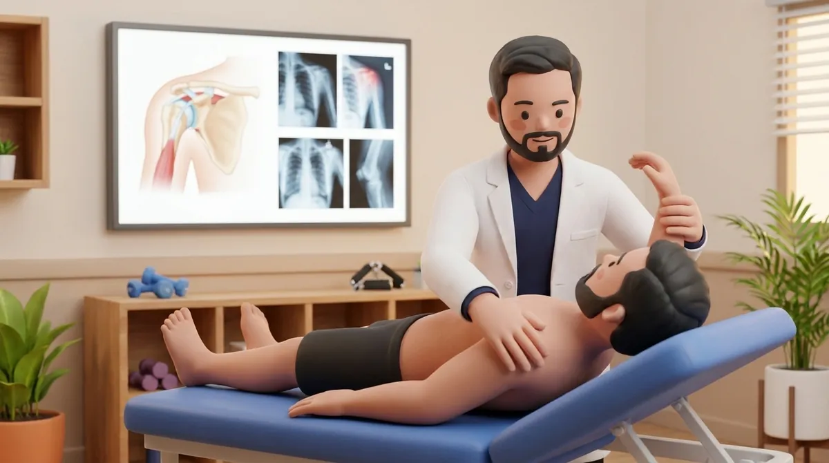
The LCL complex consists of four distinct components: the radial collateral ligament (RCL), the annular ligament, the accessory collateral ligament, and the lateral ulnar collateral ligament (LUCL). The LUCL originates from the isometric center of the lateral epicondyle and arches across the radial head to insert onto the supinator crest of the ulna. Horii described the sequential progression of elbow instability: Stage 1 involves isolated LUCL disruption producing PLRI; Stage 2 involves anterior and posterior capsular disruption with incomplete dislocation; and Stage 3 involves complete humeroulnar dislocation with UCL disruption. The lateral pivot-shift test of the elbow evaluates PLRI. With the patient supine, the examiner flexes the shoulder overhead, applies an axial load and valgus stress combined with forearm supination, and slowly flexes the elbow from full extension. At approximately forty degrees of flexion, the radial head subluxates posterolaterally, reducing with a visible clunk as flexion approaches ninety degrees.
Non-Operative Dynamic Stabilization, Pronation Protection, and LUCL Rehab
Conservative physiotherapy is indicated for low-grade partial LUCL tears or patients unable to undergo surgical reconstruction. The fundamental biomechanical principle in managing PLRI is that forearm pronation dynamically stabilizes the lateral elbow. Pronation tightens the intact medial soft tissues and engages the dynamic extensor-supinator musculature, drawing the radial head firmly against the capitellum. Active rehabilitation focuses on strengthening the extensor carpi radialis longus and brevis (ECRL/ECRB), the anconeus, and the supinator muscles. Patients are taught compensatory motion strategies, such as rising from chairs with their forearms in full pronation (palms facing inward or backward). Following surgical LUCL reconstruction using a tendon autograft, the elbow is protected in a hinged brace, restricting terminal extension and supination for six to eight weeks.
Pronated Closed-Chain Table Pushup Stabilization Drill
Objective: Strengthen dynamic lateral elbow stabilizers under axial compression while maintaining protective forearm pronation
- 1Stand facing a sturdy, waist-high wooden table with your feet placed shoulder-width apart.
- 2Place your palms flat on the tabletop with your fingers pointing inward toward each other, ensuring forearms are in full pronation.
- 3Transfer forty percent of your body weight forward onto your hands, keeping your elbows softly bent at twenty degrees.
- 4Slowly bend your elbows to thirty degrees of flexion, lowering your chest five centimeters toward the table under strict control.
- 5Push firmly through your palms to return to the twenty-degree starting position, maintaining complete forearm pronation throughout.
| Instability Stage | Disrupted Ligamentous Structures | Clinical Presentation and Physical Examination Signs |
|---|---|---|
| Stage 1: PLRI | Disruption of the lateral ulnar collateral ligament (LUCL); anterior and posterior capsular fibers intact | Positive posterolateral rotatory drawer test; positive lateral pivot-shift apprehension; snapping when rising from a chair |
| Stage 2: Incomplete Dislocation | LUCL disruption propagating through anterior and posterior capsule; coronoid rests perched on trochlea | Severe joint laxity under combined varus and supination; prominent posterior step-off; pain on terminal extension |
| Stage 3A: Complete Dislocation (Posterior) | Complete disruption of all lateral and capsular restraints; anterior bundle of UCL remains partially intact | Frank elbow dislocation requiring manual reduction; joint remains stable to valgus stress in 90 degrees flexion |
| Stage 3B: Gross Instability | Complete disruption of both LCL complex and entire UCL (anterior and posterior bundles) | Gross multidirectional instability; joint redislocates spontaneously upon release of traction; requires external fixator or dual repair |
In an awake patient, the lateral pivot-shift test is frequently negative because conscious muscle guarding and apprehension prevent the radial head from subluxating. A patient experiencing severe apprehension, muscle guarding, or pain during the maneuver confirms a positive test without needing to force an overt mechanical clunk.
Frequently Asked Questions
Why does my elbow feel like it slips out of place when I push up from a chair?
When you push up from an armrest with your hands turned outward in supination, you place body weight, varus torque, and twisting forces across your elbow. If your lateral collateral ligament is torn, this specific combination causes the radial head to slide backward out of its socket, creating a sudden catching or slipping sensation.
Do I need a doctor referral to see an elbow physiotherapist on BookPhysio.in?
BookPhysio.in does not require a physician prescription. Under the National Commission for Allied and Healthcare Professions (NCAHP) Act 2021, patients across India enjoy direct access to autonomous registered physiotherapists. Through BookPhysio.in, you connect directly with verified clinicians and pay per session with transparent fees, completely free from advance package commitments, commission markups, or platform subscription charges.
Related recovery guides & articles
BookPhysio.in Editorial Team
The BookPhysio.in editorial team writes and maintains this content, checking it against reliable clinical sources. Content follows our medical review policy: general information only, and no page claims a clinical review until a named registered physiotherapist has signed it off.
Read our medical review policy