
Medial Calcaneal Nerve Entrapment and Baxter Nerve: Differential Diagnosis in Heel Pain
In short
Learn how to distinguish Baxter nerve entrapment and medial calcaneal neuropathy from plantar fasciopathy, recognize abductor atrophy, and treat neural heel pain.
In this guide (4 sections)
While chronic plantar fasciopathy accounts for the overwhelming majority of inferior heel pain, clinicians must recognize that approximately ten to twenty percent of recalcitrant heel pain cases involve entrapment or irritation of local peripheral nerve branches. The two most critical neural structures implicated in inferior and medial heel pain are the medial calcaneal nerve and the first branch of the lateral plantar nerve, classically termed the Baxter nerve (named after Donald E. Baxter). Because the pain location of Baxter neuropathy overlaps almost identically with the medial calcaneal tuberosity insertion of the plantar fascia, it is frequently misdiagnosed as refractory plantar fasciitis. Patients subjected to months of standard fascial stretches, shockwave therapy, or night splints often experience zero relief or even symptom aggravation when an unrecognized neural compression is the primary driver of their discomfort. Accurate clinical diagnosis requires precise anatomical palpation, neurodynamic testing, and identification of characteristic motor denervation changes.
Barefoot Marble Floor Living, Prolonged Kitchen Standing, and Thin Flat Slippers
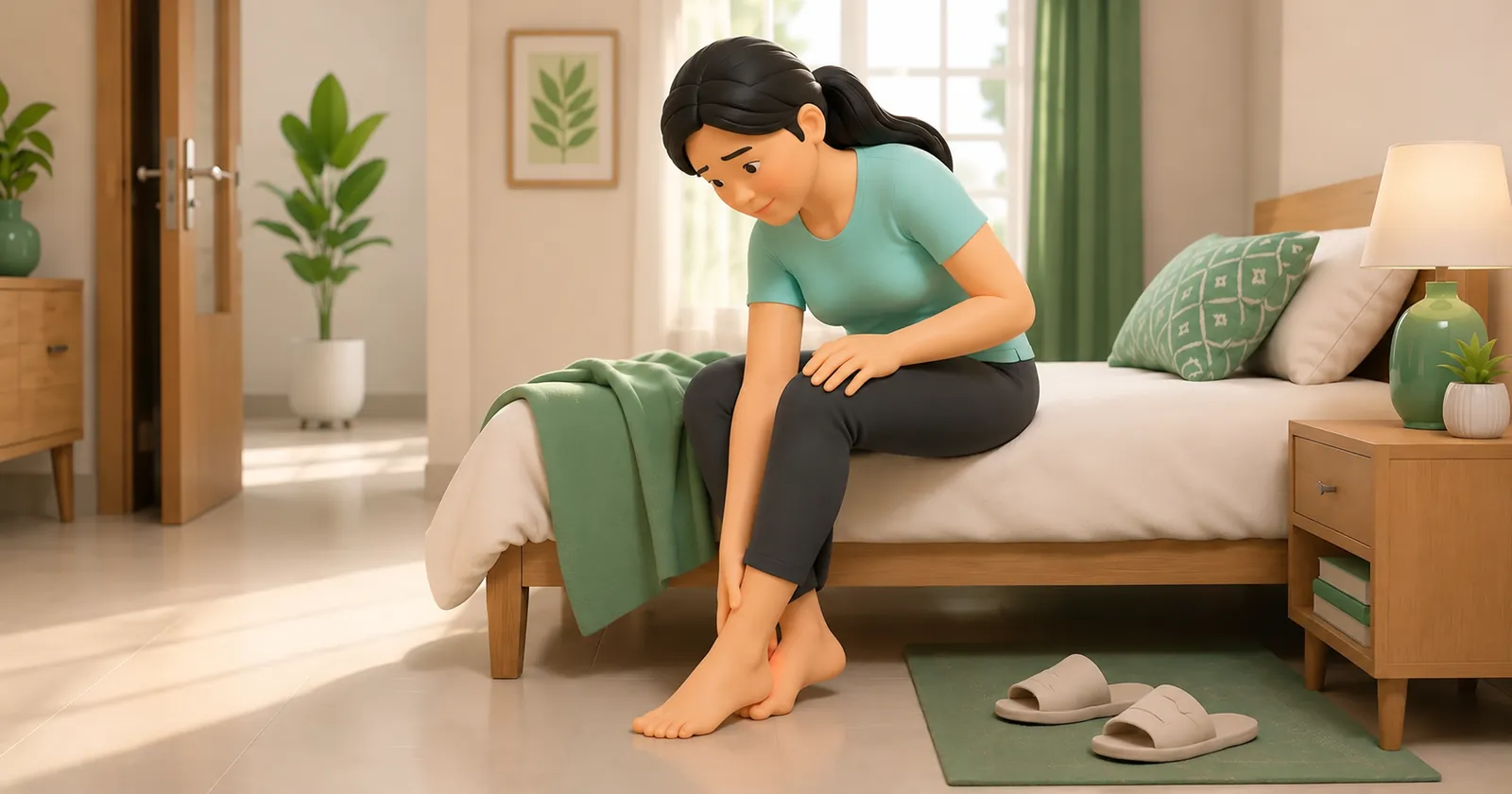
In Indian households, domestic biomechanics and traditional footwear habits significantly contribute to peripheral nerve entrapment around the heel. Polished marble and granite flooring create an unforgiving, stiff surface that generates elevated peak impact forces upon initial heel strike. Walking barefoot across these unyielding surfaces compresses the subcutaneous fat pad, directly transmitting mechanical pressure into the superficial branches of the medial calcaneal nerve. Furthermore, wearing traditional flat footwear like Kolhapuri chappals or flimsy flat rubber slippers allows the medial longitudinal arch to collapse completely during prolonged standing. When an Indian homemaker stands for three to four hours cooking at the kitchen counter with an unsupported, pronated foot, the deep fascia of the abductor hallucis muscle tightens, compressing the Baxter nerve against the medial calcaneal tuberosity and triggering burning, tingling heel pain.
Windlass Mechanism Strain, Calcaneal Enthesopathy, and Plantar Thickness
Anatomically, the Baxter nerve arises from the lateral plantar nerve near the bifurcation of the posterior tibial nerve beneath the flexor retinaculum. It travels vertically downward between the deep fascia of the abductor hallucis muscle and the medial belly of the quadratus plantae, then turns sharply laterally across the anterior margin of the medial calcaneal tuberosity to innervate the abductor digiti minimi muscle and provide sensory fibers to the calcaneal periosteum. Entrapment typically occurs at two specific anatomical pinch points: where the nerve passes beneath the rigid, deep fascial margin of the abductor hallucis, or where it passes adjacent to the medial calcaneal tuberosity (often near a traction spur). Chronic compression leads to microvascular ischemia of the endoneurium, local nerve edema, and demyelination. In chronic cases, denervation of the abductor digiti minimi muscle ensues, which can be visualized on MRI as distinct muscle fatty atrophy and high T2-weighted signal intensity.
Rathleff High-Load Towel Training, Low-Dye Taping, and Intrinsic Foot Doming
Managing neural heel pain requires a distinctly different rehabilitation approach than purely mechanical fascial loading. Aggressive tensile stretching or deep transverse friction massage, which benefits fascial enthesopathy, frequently inflames compressed neural tissue and must be avoided. Physiotherapists utilize neurodynamic mobilization techniques, performing gentle sliding and gliding maneuvers along the tibial and lateral plantar nerve tracts to restore normal intraneural excursion and reduce perineural tethering. Soft-tissue release directed specifically at the tight myofascial band of the abductor hallucis relieves mechanical pressure at the proximal entrapment site. Low-Dye taping or custom orthotics featuring a medial arch post and deep heel cup prevent excessive foot pronation, effectively widening the fibro-osseous tunnel through which the nerve travels. As neural irritability subsides, intrinsic foot muscle doming and progressive calf strengthening are carefully introduced.
Tibial and Plantar Nerve Neurodynamic Slider Mobilization
Objective: Promote longitudinal intraneural excursion and relieve perineural mechanical tension along the tibial and Baxter nerve tracts
- 1Sit comfortably on a treatment table or firm bed with your legs extended straight forward and your torso upright.
- 2Begin by slowly pointing your ankle downward (plantarflexion) and turning your foot inward while simultaneously bending your head and neck forward (chin to chest).
- 3Next, smoothly reverse the motion: flex your ankle upward toward your shin (dorsiflexion) and turn your foot outward while simultaneously extending your head to look up toward the ceiling.
- 4Continue alternating smoothly between these two coordinated positions in a rhythmic, continuous gliding motion without holding end-range tension.
- 5Ensure the exercise produces only a gentle stretching or pulling sensation without provoking sharp, shooting electrical pain.
| Diagnostic Feature | Plantar Fasciopathy | Baxter Nerve Entrapment (First Branch Lateral Plantar) | Clinical Diagnostic Action |
|---|---|---|---|
| Pain Quality & Pattern | Sharp, stabbing mechanical pain worst on first morning steps; eases with warm-up | Burning, tingling, or radiating ache; worsens progressively as the day proceeds | Evaluate symptom trajectory and response to prolonged static standing |
| Palpation Site | Focal point tenderness directly on the medial calcaneal tuberosity | Maximal tenderness 1 to 2 cm anterior and distal to the medial calcaneal tuberosity | Perform Tinel percussion test over the abductor hallucis fascial margin |
| Diagnostic Imaging Findings | Ultrasound demonstrates fascial thickness >4.0 mm and loss of fibrillar pattern | MRI reveals fatty atrophy and denervation edema in the abductor digiti minimi | Order dedicated foot MRI if symptoms fail to respond to 6 weeks of loading |
If a patient presents with rapid motor weakness in the foot, sensory loss extending across the entire plantar aspect, or radiating pain that extends upward into the calf along the sciatic distribution, a proximal lumbosacral radiculopathy (L5-S1) or systemic peripheral polyneuropathy must be considered. Furthermore, severe localized bone tenderness to calcaneal squeezing mandates immediate exclusion of a calcaneal stress fracture. Refer for prompt orthopaedic and neurological evaluation.
Frequently Asked Questions
How do I know if my heel pain is a pinched nerve rather than regular plantar fasciitis?
While regular plantar fasciitis characteristically causes severe pain during the very first steps in the morning that eases after walking, Baxter nerve entrapment typically produces a burning, tingling, or throbbing ache that intensifies as you stay on your feet throughout the day. You may also feel pain radiating along the outside of your heel or into your smaller toes. A registered physiotherapist can perform specific neurological tests, such as Tinel percussion and neurodynamic tension tests, to accurately distinguish between the two conditions.
Do I need a doctor referral to treat plantar fasciitis on BookPhysio.in?
BookPhysio.in does not require a doctor referral. Under the National Commission for Allied and Healthcare Professions (NCAHP) Act 2021, patients across India enjoy direct access to autonomous registered physiotherapists. Through BookPhysio.in, you connect directly with verified clinicians and pay per session with transparent fees, completely free from advance package commitments, commission markups, or platform subscription charges.
Related recovery guides & articles
BookPhysio.in Editorial Team
The BookPhysio.in editorial team writes and maintains this content, checking it against reliable clinical sources. Content follows our medical review policy: general information only, and no page claims a clinical review until a named registered physiotherapist has signed it off.
Read our medical review policy