
Patellar Mobilization and Anterior Interval Scar Release Techniques
In short
Master manual therapy techniques for patellar glides and anterior interval scar release to eliminate arthrofibrosis following meniscal surgery.
In this guide (4 sections)
Arthrofibrosis and soft-tissue scar tethering represent some of the most frustrating and disabling complications encountered following knee trauma, arthroscopic meniscal repair, or prolonged post-operative immobilization. During arthroscopic surgery, surgical instrumentation, portal incisions, and fluid distension traumatize the infrapatellar fat pad (Hoffa fat pad) and anterior capsule. Without timely manual intervention, dense fibrous scar tissue forms within the anterior interval, the anatomical space between the posterior surface of the patellar tendon, the infrapatellar fat pad, and the anterior surface of the proximal tibia. Scar contracture in this space tethers the patellar tendon firmly to the tibia, blocking normal patellar superior and inferior translation. This patellar entrapment severely restricts both terminal knee extension and deep flexion, producing chronic anterior knee pain and abnormal joint mechanics.
Indian Post-Op Realities: Traditional Immobilization and Fear of Movement
In Indian domestic culture, post-operative care is often dominated by traditional beliefs that favor prolonged complete bed rest and absolute joint immobility. Well-meaning family members frequently insist that a patient recovering from meniscal surgery remain completely motionless in bed for several weeks, warning that any knee movement might disrupt surgical stitches. In the absence of regular active range of motion and manual therapy, dense collagenous adhesions rapidly cement the patella to the anterior tibia. When the patient finally attempts to bend the knee to manage personal hygiene or sit on a chair, the kneecap is completely locked, producing agonizing anterior pain. Educating families on the critical necessity of gentle early patellar mobilizations prevents arthrofibrotic joint freezing.
Anterior Interval Anatomy, Patellar Kinematics, and Fibrotic Tethering
Normal knee kinematics depend entirely on the free, unhindered excursion of the patella within the trochlear groove. During knee flexion from 0 to 90 degrees, the patella must glide inferiorly by approximately 1.5 to 2 centimetres and tilt to maintain congruency. Conversely, during terminal knee extension, the quadriceps pulls the patella superiorly into its resting supratrochlear pouch. In a healthy joint, the anterior interval is lined with supple, lubricated synovial folds and compliant adipose tissue. When inflamed by surgical trauma or hemarthrosis, fibroblasts proliferate and deposit disorganized Type III collagen, obliterating the anterior interval. This tethering anchors the patellar tendon, creating patella baja (inferior patellar displacement). As the patient attempts to flex the knee, the trapped patellar tendon cannot elongate, creating an unyielding mechanical block and crushing the sensitive infrapatellar fat pad.
Manual Therapy Protocols: Multiplanar Patellar Glides and Soft-Tissue Mobilization
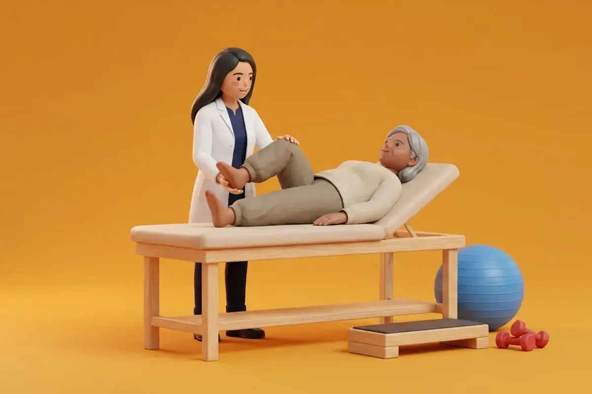
Restoring anterior joint compliance requires systematic manual therapy initiated in the earliest post-operative phase. With the patient supine and the knee supported in full extension with quadriceps completely relaxed, the physiotherapist applies gentle, sustained manual glides in four distinct directions: superior, inferior, medial, and lateral. Superior patellar glides are prioritized to restore knee extension, while inferior glides restore knee flexion. Medial and lateral glides stretch tight retinacular tissues. To release anterior interval scarring directly, the clinician performs gentle transverse friction massage across the infrapatellar tendon margins, followed by gentle posterior tilting of the inferior pole to mobilize the deep fat pad. Patients are thoroughly instructed on performing self-mobilization techniques at home multiple times daily.
Patient-Administered Superior and Inferior Patellar Self-Glides
Objective: Maintain normal patellar mobility and prevent anterior interval scar tethering during early post-operative recovery
- 1Sit comfortably on a bed or flat floor with your affected leg extended completely straight and your thigh muscles fully relaxed.
- 2Place the pads of your thumbs along the inferior (bottom) curved border of your kneecap.
- 3Gently push the kneecap upward toward your hip until you feel a firm, comfortable resistance, holding for 3 seconds.
- 4Next, place the index fingers or heels of your hands along the superior (top) border of your kneecap.
- 5Gently push the kneecap downward toward your foot until firm resistance is reached, holding for 3 seconds.
- 6Ensure your leg remains completely relaxed throughout the glides, feeling the kneecap slide smoothly within its groove.
| Mobilization Vector | Manual Technique & Direction | Targeted Anatomical Structure | Restored Knee Motion | Clinical Indication |
|---|---|---|---|---|
| Superior Patellar Glide | Push kneecap upward toward hip with thumbs | Infrapatellar tendon & fat pad posterior interface | Full terminal knee extension (0 degrees) | Extension lag, patella baja, post-repair Weeks 1 to 6 |
| Inferior Patellar Glide | Push kneecap downward toward foot with web-space | Quadriceps tendon & suprapatellar pouch | Knee flexion beyond 90 degrees | Flexion stiffness, subacute rehabilitation |
| Medial Patellar Glide | Push kneecap inward toward opposite leg | Lateral retinaculum & iliotibial band expansions | Reduces patellar lateral tilt and tilt-related friction | Patellofemoral compression syndrome, lateral tightness |
| Anterior Interval Release | Deep soft-tissue transverse friction behind tendon | Hoffa fat pad adhesions to anterior tibial cortex | Both terminal extension and deep flexion | Arthrofibrosis, chronic post-surgical joint stiffness |
Aggressive, forceful manual pinching of the infrapatellar fat pad can provoke severe secondary inflammation termed Hoffa disease (hoffitis). If a patient develops acute, burning pain directly behind the patellar tendon, accompanied by prominent swelling that bulges on either side of the tendon and exquisite tenderness to light pinch during terminal extension (positive Hoffa sign), manual therapy must be softened immediately, cryotherapy applied, and hyperextension taped to allow the inflamed fat pad to settle.
Frequently Asked Questions
Why does my kneecap feel glued down after knee surgery?
During surgery and early recovery, fluid swelling and natural healing create sticky fibrin bands around your kneecap and the fat pad beneath it. If your kneecap is not moved regularly with gentle manual glides, these bands harden into thick fibrous scar tissue that glues your kneecap down. Performing gentle patellar self-glides keeps the joint tissues flexible and prevents joint freezing.
Do I need a doctor referral to treat a meniscal tear on BookPhysio.in?
BookPhysio.in does not require a doctor referral. Under the National Commission for Allied and Healthcare Professions (NCAHP) Act 2021, patients across India enjoy direct access to autonomous registered physiotherapists. Through BookPhysio.in, you connect directly with verified clinicians and pay per session with transparent fees, completely free from advance package commitments, commission markups, or platform subscription charges.
Related recovery guides & articles
BookPhysio.in Editorial Team
The BookPhysio.in editorial team writes and maintains this content, checking it against reliable clinical sources. Content follows our medical review policy: general information only, and no page claims a clinical review until a named registered physiotherapist has signed it off.
Read our medical review policy