
Postoperative Atelectasis: Micro-Atelectasis to Lobar Collapse Progression
In short
Understand how postoperative micro-atelectasis progresses to full lobar collapse. Learn alveolar recruitment mechanics, surfactant preservation, and physio protocols.
In this guide (2 sections)
Postoperative atelectasis is the most prevalent respiratory complication encountered after major abdominal and thoracoabdominal operations, affecting up to ninety percent of patients under general anesthesia. What begins as asymptomatic subclinical micro-atelectasis in dependent lung segments can rapidly progress into segmental or complete lobar collapse if hypoventilation, airway clearance failure, and pain-induced shallow breathing persist. Recognizing the pathophysiological cascade from microscopic alveolar collapse to consolidated lobar collapse is critical for implementing timely, restorative chest physiotherapy.
Surgical Pathophysiology and Thoracoabdominal Mechanics
The genesis of postoperative atelectasis involves three synergistic mechanisms: compression atelectasis, absorption atelectasis, and surfactant displacement. Under general anesthesia, muscle relaxants reduce diaphragmatic tone, permitting the heavy abdominal contents to shift cephalad, compressing dependent basal alveoli. Administration of high fractions of inspired oxygen (FiO2) washes out nitrogen, the structural gas that maintains alveolar patency; when alveolar oxygen rapidly diffuses into pulmonary capillaries faster than inspired gas replaces it, complete alveolar collapse occurs. As surfactant molecules migrate out of collapsed alveoli, critical opening pressures rise dramatically, requiring higher transpulmonary pressures to reopen lung units and driving a steep increase in intrapulmonary right-to-left shunt.
| Stage | Alveolar Physiology | Clinical Signs and Auscultation | Physiotherapy Action Plan | |
|---|---|---|---|---|
| Stage 1: Micro-Atelectasis | Scattered alveolar collapse in dependent lung bases; loss of FRC | Mild SpO2 decline (92-94% room air); faint fine end-inspiratory crackles | Frequent hourly deep breathing with 3-second holds, upright sitting posture | |
| Stage 2: Segmental Collapse | Confluent collapse of multiple bronchopulmonary segments | Tachypnea (RR > 22), localized dullness to percussion, decreased breath sounds | Active Cycle of Breathing Techniques (ACBT), PEP bottle therapy, early ambulation | |
| Stage 3: Lobar Collapse | Complete obstruction and collapse of entire anatomical lobe | Severe hypoxemia, bronchial breathing, tracheal shift, high fever, purulent sputum | Intensive postural drainage, chest percussion, assisted huffing, potential bronchoscopy |
Alveolar Recruitment Protocols and Bedside Management
Reversing atelectasis requires generating sufficient transpulmonary pressure gradients to overcome critical opening thresholds. The physiological foundation is sustained maximal inspiration (SMI) paired with an end-inspiratory pause. During the pause, gas redistributes from well-ventilated units into collapsed adjacent alveoli via collateral channels, specifically the interalveolar pores of Kohn and bronchoalveolar canals of Lambert. This collateral recruitment gently re-inflates collapsed zones without causing barotrauma.
Sustained Maximal Inspiration with Collateral Recruitment Hold
Objective: Re-expand collapsed basal alveoli and enhance collateral ventilation throughout dependent lung tissue.
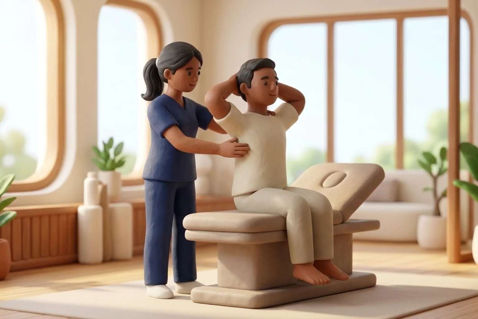
- 1Elevate the head of the bed to at least 45 degrees, or ideally assist the patient into an upright bedside chair.
- 2Place both hands firmly on the lower lateral ribs to provide tactile feedback.
- 3Instruct the patient to exhale completely and gently through the mouth until lungs feel empty.
- 4Guide a slow, deep, maximal inhalation through the nose, expanding the lower chest against hand pressure.
- 5Instruct the patient to hold the breath at peak lung volume for a full 3 to 5 seconds before releasing a passive, relaxed exhalation.
Preventing Post-Discharge Respiratory Deterioration at Home in India
In Indian home settings, elderly patients often remain confined to bed following major surgery, mistakenly believing total bed rest speeds recovery. This prolonged immobility in recumbent positions accelerates dependent atelectasis, especially during cool winter months or in air-conditioned rooms with poor air circulation. Caregivers must be educated that daily sitting in a firm armchair for meals, walking through the corridor or courtyard, and keeping windows open for fresh ventilation are vital measures to keep lung bases clear.
Long-Term Recovery Trajectory and Milestones
Most uncomplicated atelectasis resolves within five to seven days when early mobilization and deep breathing protocols are sustained. Continued daily walking, volumetric incentive spirometer practice, and gradual posture re-alignment ensure long-term preservation of functional residual capacity and prevent secondary hypostatic pneumonia.
Atelectasis within the first forty-eight hours is the leading cause of early postoperative pyrexia. Notify the medical team if temperature exceeds 38.0 degrees Celsius, accompanied by sudden SpO2 drops or absent breath sounds on auscultation.
Why does high oxygen delivery sometimes worsen postoperative atelectasis?
Breathing one hundred percent oxygen removes nitrogen from the alveoli. Because oxygen is rapidly absorbed into the bloodstream across alveolar membranes, gas volume within poorly ventilated alveoli drops rapidly to zero, causing complete absorption collapse. Maintaining the lowest effective FiO2 preserves alveolar nitrogen and prevents this collapse.
Do I need a doctor referral to consult a home physiotherapist in India?
Under the National Commission for Allied and Healthcare Professions (NCAHP) Act 2021, patients in India enjoy direct access to registered physiotherapists without requiring a mandatory doctor referral. At BookPhysio.in, you consult verified physiotherapists and pay directly per session without hidden fees or platform commissions.
Related recovery guides & articles
BookPhysio.in Editorial Team
The BookPhysio.in editorial team writes and maintains this content, checking it against reliable clinical sources. Content follows our medical review policy: general information only, and no page claims a clinical review until a named registered physiotherapist has signed it off.
Read our medical review policy