
Scapular Dyskinesis Correction in Chronic Rotator Cuff Deficiency
In short
Abnormal scapular motion alters glenoid orientation and narrows the subacromial space. Learn how physiotherapists classify and correct scapular dyskinesis.
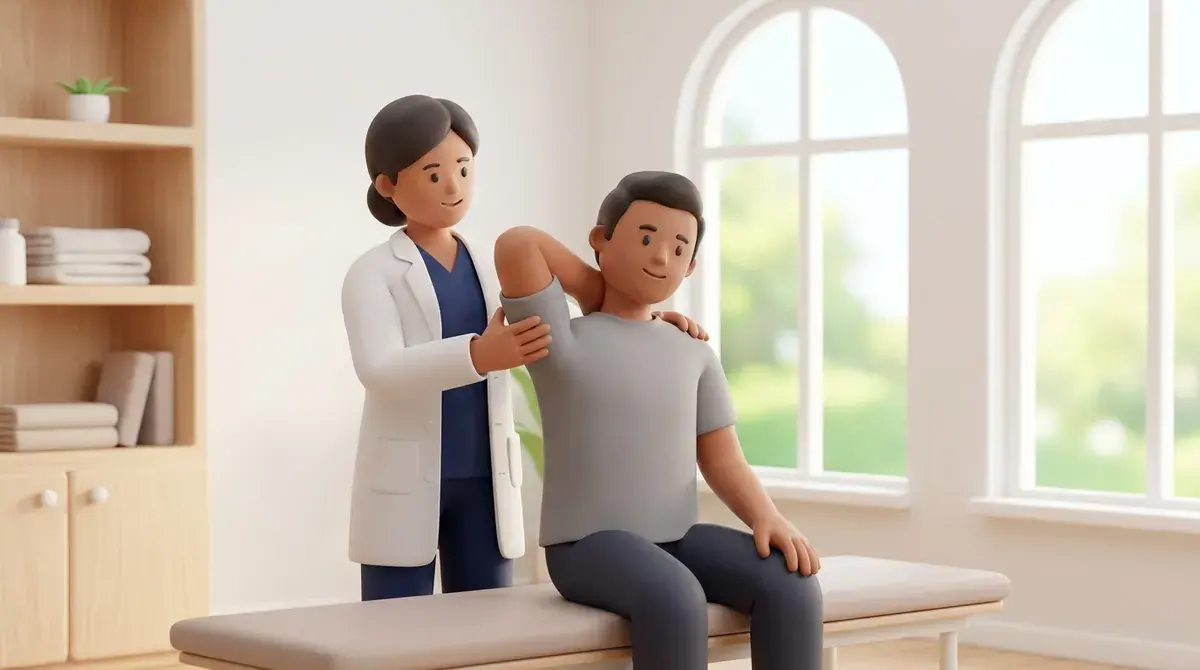
In this guide (4 sections)
The scapula serves as the mobile foundation for the entire upper extremity, linking the axial skeleton to the humerus via the clavicular strut. In healthy movement, the scapula moves in dynamic synchrony with the humerus, a relationship known as scapulohumeral rhythm. In chronic rotator cuff deficiency, this harmonious movement is frequently disrupted, giving rise to scapular dyskinesis. Defined as observable alterations in normal resting scapular position or dynamic motion during arm elevation and lowering, dyskinesis alters the orientation of the glenoid fossa. This maladaptive movement narrows the subacromial space, increases tensile shear across torn or repairing cuff tendons, and exacerbates secondary impingement. Correcting dyskinesis is a core element of both conservative management and post-surgical rehabilitation.
Impact of Abnormal Scapular Motion on Daily Life in Indian Households
In Indian domestic environments, scapular dyskinesis manifests as visible shoulder asymmetry and functional weakness during common tasks. When a homemaker reaches forward to turn on a kitchen exhaust fan or wipe down kitchen tiles, family members often notice the shoulder blade winging outward like a small wing. Carrying heavy domestic shopping bags from local vegetable markets causes the affected shoulder to sag downward, triggering persistent aching along the medial border of the shoulder blade and base of the neck. In older adults who spend hours sewing, cutting fabrics, or reading religious scriptures while seated on the floor with a rounded back, poor thoracic posture directly locks the scapula in anterior tilt. Physiotherapists evaluate these postural habits and prescribe functional movement retraining.
Kibler Classification Patterns and the Serratus-Trapezius Force Couple
W. Ben Kibler classified scapular dyskinesis into three distinct clinical patterns based on prominent anatomical landmarks: Type I (inferior medial angle prominence, indicating anterior tipping); Type II (entire medial border prominence; this indicates excessive internal rotation or true winging); and Type III (superior border prominence with shoulder hiking, signifying excessive upper trapezius overactivity). Biomechanically, normal arm elevation requires three coordinated scapular motions: upward rotation (sixty degrees), posterior tilt (twenty to thirty degrees), and external rotation. These movements are executed by the scapulothoracic force couple: the upper trapezius initiates early elevation, the serratus anterior drives upward rotation and pins the medial border against the thoracic wall, and the lower trapezius provides posterior tilt and inferior depression. When the serratus anterior and lower trapezius become weak, dyskinesis inevitably develops.
Neuromuscular Retraining: Muscle Activation and Pectoralis Minor Lengthening
Clinical management of scapular dyskinesis follows a systematic two-pronged approach: releasing tight anterior structures and activating inhibited posterior stabilizers. Physiotherapists perform manual release and guided self-stretching of the pectoralis minor, whose adaptive shortening pulls the coracoid process anteriorly and locks the scapula in anterior tilt. Concurrently, therapeutic exercise focuses on conscious scapular setting and strengthening of the serratus anterior and lower trapezius. Low-load motor control drills, such as the Scapular Assistance Test (SAT) and Scapular Retraction Test (SRT), demonstrate immediate pain relief when normal kinematics are manually restored, confirming the clinical diagnosis and guiding exercise prescription.
Corner Pectoralis Minor Stretch and Lower Trapezius Squeeze
Objective: Elongate the tight pectoralis minor muscle to restore posterior scapular tilt and increase subacromial space
- 1Stand facing the corner of a room, approximately two feet away from the walls, with your feet hip-width apart.
- 2Place your forearms flat against the adjacent walls at shoulder height, keeping your elbows bent to ninety degrees.
- 3Step one foot slightly forward and gently lean your chest toward the corner until you feel a comfortable stretch across your chest.
- 4While maintaining the stretch, gently squeeze your lower shoulder blades downward and together for 5 seconds.
- 5Step back smoothly to the starting position, relax for 3 seconds, and repeat with calm, continuous breathing.
| Dyskinesis Pattern | Prominent Landmark & Kinematic Abnormality | Targeted Muscle Imbalance & Corrective Exercise Focus |
|---|---|---|
| Type I: Inferior Angle Prominence | Inferior medial angle projects away from ribcage; excessive anterior tilt of scapula during elevation. | Tight pectoralis minor; weak lower trapezius and serratus anterior; corner stretch and prone Y-raises. |
| Type II: Medial Border Prominence | Entire medial border lifts off thoracic wall; excessive internal rotation; classical dynamic winging. | Severe serratus anterior weakness or long thoracic nerve neuropraxia; wall push-up plus and dynamic hug drills. |
| Type III: Superior Border Prominence | Superior angle elevates prematurely; excessive early shoulder hiking and lack of true upward rotation. | Hyperactive upper trapezius and levator scapulae; inhibited lower trapezius; conscious shrug-suppression drills. |
| Scapular Assistance Test (SAT) | Examiner manually assists upward rotation and posterior tilt during active forward elevation. | Immediate reduction in painful arc confirms dyskinesis as primary contributor to impingement symptoms. |
Gross, unresolving medial border winging (Type II dyskinesis) that fails to improve with exercise may represent true serratus anterior denervation resulting from a long thoracic nerve traction injury. Electrodiagnostic studies (EMG/NCS) are indicated if severe winging is accompanied by profound overhead reaching weakness.
Frequently Asked Questions
Can correcting my shoulder blade posture help my rotator cuff pain go away?
Yes. Restoring normal shoulder blade movement tilts the roof of your shoulder (the acromion) upward and backward during arm lifting. This dynamic clearance expands the subacromial space, taking compressive pressure off the rotator cuff tendons and significantly reducing pain during daily reaching activities.
Do I need a doctor referral to see a rotator cuff physiotherapist on BookPhysio.in?
BookPhysio.in does not require a physician prescription. Under the National Commission for Allied and Healthcare Professions (NCAHP) Act 2021, patients across India enjoy direct access to autonomous registered physiotherapists. Through BookPhysio.in, you connect directly with verified clinicians and pay per session with transparent fees, completely free from advance package commitments, commission markups, or platform subscription charges.
Related recovery guides & articles
BookPhysio.in Editorial Team
The BookPhysio.in editorial team writes and maintains this content, checking it against reliable clinical sources. Content follows our medical review policy: general information only, and no page claims a clinical review until a named registered physiotherapist has signed it off.
Read our medical review policy