
Tibial Tubercle Osteotomy (TTO): Fulkerson Realignment, Rehabilitation Protocols, and Phasing
In short
Understand surgical realignment of the extensor mechanism through tibial tubercle osteotomy and learn phased rehabilitation protocols for bone union.
In this guide (4 sections)
Tibial tubercle osteotomy (TTO) represents a powerful, definitive surgical realignment procedure indicated for complex patellofemoral disorders that fail exhaustive conservative therapy. In individuals exhibiting severe patellar maltracking, recurrent patellar instability, or localized distal and lateral patellofemoral cartilage degradation, underlying anatomical deformities are frequently identified. These include an excessively lateralized tibial tuberosity (quantified on cross-sectional CT or MRI as an elevated tibial tubercle-trochlear groove distance, TT-TG, exceeding twenty millimetres) or patella alta (an abnormally elevated patella quantified by a Caton-Deschamps index exceeding 1.2). The classic Fulkerson osteotomy involves an oblique, anteromedialization (AMZ) osteotomy cut of the tibial tuberosity, which is surgically shifted medially to normalize the Q-angle and anteriorly to decompress retropatellar contact stress, secured with rigid bicortical screws. Because TTO involves creating a controlled surgical bone fracture, rehabilitation must rigorously respect osseous healing timelines.
Complex Orthopaedic Reconstructions, Home Mobility, and Phased Healing in India
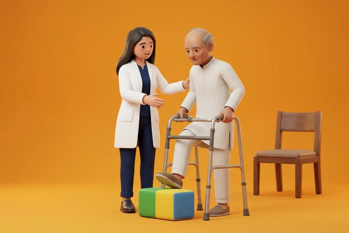
Undergoing a major realignment procedure like a Fulkerson osteotomy represents a significant life event for Indian patients. Following discharge from orthopaedic specialty hospitals in cities like Delhi, Mumbai, Bengaluru, or Ahmedabad, patients return to multi-generational households that often feature floor-level living, low sofas, or flights of entrance stairs. The immediate domestic challenge is navigating home mobility while strictly adhering to non-weight-bearing restrictions. Patients must be educated on utilizing crutches safely, avoiding sudden twisting movements, and implementing ergonomic bed and seating modifications. Close coordination between the operating orthopaedic surgeon and the treating physiotherapist ensures that the bony osteotomy heals solidly before aggressive weight-bearing loads are introduced.
Osteotomy Biomechanics, Contact Stress Redistribution, and Bone Consolidation
The mechanical elegance of an anteromedial tibial tubercle osteotomy lies in its simultaneous two-plane realignment. Medial translation of the tubercle (typically eight to ten millimetres) reduces the lateral vector force of the quadriceps extensor mechanism, normalizing the TT-TG distance to physiological levels (ten to twelve millimetres) and re-centering the patella within the trochlear groove. Concurrently, anterior translation (typically five to eight millimetres) increases the mechanical extensor lever arm, lifting the distal and lateral patellar facets away from the femoral condyles and reducing peak retropatellar contact stress by up to thirty-five percent. However, the osteotomy site relies on secondary bone healing through cortical bridging and trabecular consolidation. Premature high-load quadriceps contraction or full unbraced weight-bearing risks catastrophic bone non-union, osteotomy displacement, or screw pull-out.
Clinical Protocols: Non-Weight-Bearing Protection to Radiographic Bone Union
Rehabilitation following TTO is structured into four milestone-driven phases. Phase 1 (Weeks 0 to 6) is the bone protection phase: the patient is strictly non-weight-bearing to partial weight-bearing (twenty-five percent body weight) using bilateral axillary or elbow crutches, with a hinged knee brace locked in full extension for ambulation. Passive range of motion is permitted in safe zones (typically zero to sixty degrees in Weeks 1-3, progressing to ninety degrees by Week 6). Quadriceps setting and straight leg raises are performed strictly with the brace locked. Phase 2 (Weeks 7 to 12) begins upon radiographic confirmation of bone healing: the patient progresses to full weight-bearing, weans the brace, and restores full knee flexion. Phase 3 (Months 3 to 6) rebuilds kinetic chain strength, and Phase 4 (Months 6+) guides return to impact loading.
Brace-Locked Isometric Straight Leg Raise for Extensor Activation
Objective: Maintain quadriceps motor unit recruitment and prevent disuse atrophy while protecting the healing tibial osteotomy
- 1Lie flat on your back on a firm plinth or mattress, ensuring your post-operative hinged brace is securely fastened and locked at 0 degrees extension.
- 2Bend your non-operated knee to 90 degrees with your foot flat on the bed for pelvic stability.
- 3Firmly tighten your operated thigh muscles, pressing the back of your knee downward into the brace.
- 4Keeping the knee completely locked in full extension, slowly lift your operated leg 12 inches off the mattress over 2 seconds.
- 5Hold the leg elevated steadily for 3 to 5 seconds without allowing your knee to bend or sag inside the brace, then slowly lower.
- 6Relax your thigh completely for 3 seconds before initiating the next repetition.
| Rehabilitation Phase | Post-Operative Timeline | Weight-Bearing & Bracing Restrictions | Key Clinical Milestones & Interventions |
|---|---|---|---|
| Phase 1: Bone Union Protection | Weeks 0 to 6 post-surgery | Non-to-partial weight bearing on crutches; brace locked at 0 degrees | Passive ROM 0 to 90 degrees; brace-locked quad sets and leg raises |
| Phase 2: Progressive Loading | Weeks 7 to 12 post-surgery | Progress to full weight-bearing after radiographic union confirmed | Wean crutches and brace; achieve full flexion; stationary cycling |
| Phase 3: Kinetic Chain Strengthening | Months 3 to 6 post-surgery | Full unbraced ambulation; symmetrical gait mechanics | Leg press, shallow squats (0-45 deg), step-downs, gluteal loading |
| Phase 4: Functional Impact Return | Months 6 to 9+ post-surgery | Full athletic and domestic clearance; unrestricted movement | Graduated running progression, agility drills, sports-specific training |
If a patient recovering from a tibial tubercle osteotomy experiences a sudden slip, trip, or accidental full-weight stumble accompanied by an audible crack, severe sharp pain over the proximal tibia, and visible displacement or swelling, an acute osteotomy fracture or screw failure must be suspected. The limb must be splinted immediately in extension, made strictly non-weight-bearing, and referred for urgent orthopaedic radiographs.
Frequently Asked Questions
Why do I have to keep my brace locked straight for six whole weeks after a Fulkerson osteotomy?
A Fulkerson osteotomy involves cutting your shin bone to move the attachment of your kneecap tendon into a better position, secured with screws. In the first six weeks, your bone is actively knitting back together. Keeping your brace locked straight when walking prevents your powerful thigh muscles from pulling on the healing bone fragment, ensuring the bone heals solidly without shifting.
Do I need a doctor referral to treat patellofemoral pain on BookPhysio.in?
BookPhysio.in does not require a doctor referral. Under the National Commission for Allied and Healthcare Professions (NCAHP) Act 2021, patients across India enjoy direct access to autonomous registered physiotherapists. Through BookPhysio.in, you connect directly with verified clinicians and pay per session with transparent fees, completely free from advance package commitments, commission markups, or platform subscription charges.
Related recovery guides & articles
BookPhysio.in Editorial Team
The BookPhysio.in editorial team writes and maintains this content, checking it against reliable clinical sources. Content follows our medical review policy: general information only, and no page claims a clinical review until a named registered physiotherapist has signed it off.
Read our medical review policy