
Weight-Bearing Computed Tomography (WBCT) in Dynamic Instability
In short
Conventional supine CT scans conceal dynamic vertebral slippage. Learn how weight-bearing CT reveals hidden instability and guides precision rehabilitation.
In this guide (4 sections)
The radiological evaluation of lumbar spondylolysis and spondylolisthesis is undergoing an evolutionary paradigm shift driven by the emergence of cone-beam Weight-Bearing Computed Tomography (WBCT). For decades, advanced volumetric cross-sectional imaging of the human spine has been performed almost exclusively with the patient lying supine within conventional gantry CT or MRI scanners. However, supine positioning eliminates the primary physical force governing human spinal mechanics: gravity. In the recumbent state, vertebral bodies spontaneously reduce, foraminal dimensions expand, and dynamic instability remains completely concealed. By scanning the spine in natural erect standing postures under true physiological body weight, WBCT unmasks occult translational slippage, provides ultra-high-resolution cortical bone detail, and transforms clinical rehabilitation decision-making.
Indian Tertiary Hospital Technology, Urban Adoption, and Diagnostic Precision
In leading medical and orthopedic centers across major Indian metros, such as Mumbai, Bengaluru, Hyderabad, and Delhi, advanced weight-bearing imaging systems are rapidly gaining clinical adoption. Patients who have suffered for years with unexplained lower back pain and normal supine CT scans are finally receiving definitive diagnoses when scanned while standing upright. However, across Tier-2 cities and regional hospitals, conventional supine scanners remain the universal standard. Patients frequently travel long distances to tertiary centers after experiencing recurrent failure of conservative treatments, highlighting the importance of understanding dynamic imaging findings to guide precise, localized physiotherapy interventions.
Gravity-Induced Shear Surges, 3D Cone-Beam Physics, and Foraminal Occlusion
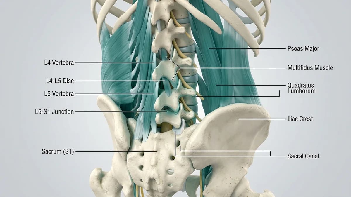
The biomechanical superiority of WBCT stems from its ability to capture true gravitational kinematics. When an individual transitions from lying flat to standing upright, axial body weight and the natural inclination of the sacral plateau generate an immediate anterior shear vector across L5-S1. In patients with isthmic or degenerative spondylolisthesis, upright WBCT demonstrates an average increase in vertebral translation of two to four millimeters compared to supine scans, frequently reclassifying Meyerding Grade I slips into Grade II. Furthermore, 3D cone-beam volumetric reconstructions reveal dynamic neuroforaminal occlusion: as the superior vertebra slides forward under gravity, the subpedicular height collapses by up to thirty-five percent, directly compressing the exiting L5 nerve root in ways completely invisible on recumbent imaging.
Image-Guided Stability Training, Upright Motor Retraining, and Progression
The quantitative insights provided by WBCT enable physiotherapists to design precision rehabilitation programs tailored to the patient actual standing biomechanics. When WBCT reveals significant occult upright translation, clinicians recognize that non-weight-bearing exercises in supine positions are insufficient to prepare the patient for upright functional life. Rehabilitation rapidly progresses from initial mat-based motor control to standing closed-chain stabilization drills. Using real-time mirrors and wearable biofeedback sensors, patients are trained to actively engage the abdominal wall and gluteal musculature in upright postures, counteracting the gravity-induced anterior shear demonstrated on their standing scans.
Standing Anti-Shear Wall Press with Multi-Planar Core Engagement
Objective: Train active upright muscular stiffness to counteract gravity-induced vertebral translation demonstrated on WBCT
- 1Stand facing a solid wall, positioned approximately two feet away, with your feet shoulder-width apart.
- 2Place both open palms flat against the wall at shoulder height, keeping your elbows slightly unlocked.
- 3Establish an upright, neutral spinal posture, engaging your abdominal muscles with a firm forty percent brace.
- 4Push firmly into the wall with your hands as if attempting to push the wall away, without leaning your body forward.
- 5Simultaneously squeeze your buttocks firmly, feeling your entire core and pelvic musculature stiffen against gravity, hold for 10 seconds.
| Imaging Parameter | Conventional Supine CT | Weight-Bearing Cone-Beam CT (WBCT) |
|---|---|---|
| Patient Scanning Posture | Horizontal supine on a flat examination couch with knees often supported on a bolster | True erect standing weight-bearing posture under full natural gravitational body mass |
| Vertebral Slip Measurement | Spontaneous reduction occurs; slip magnitude is underestimated by an average of 30-50% | Captures true functional displacement; unmasks occult translation and higher Meyerding grades |
| Neuroforaminal Dimensions | Foramina appear patent and widened due to the absence of axial compressive loading | Reveals true dynamic subpedicular narrowing and real-time nerve root entrapment |
| Radiation Exposure Dose | Standard multidetector CT radiation dose (moderate to high) | Cone-beam technology delivers significantly lower radiation dose (up to 60-70% reduction) |
When weight-bearing imaging reveals profound dynamic narrowing of the central spinal canal (cross-sectional area collapsing below 75 square millimeters upon standing) accompanied by severe bilateral leg weakness or numbness, structural stenosis is advanced. In such cases, high-velocity physiotherapy manipulations are dangerous and the patient must be referred for neurosurgical decompression consultation.
Frequently Asked Questions
Why did my standing X-ray or standing CT show a worse slip than my lying-down MRI?
Your standing scans showed a worse slip because they captured your spine under the real-world effects of gravity. When you lie flat inside an MRI tube, your body weight is completely removed from your spine, allowing your slipped vertebra to slide naturally backward into a more normal position. When you stand upright, your upper body weight pushes downward and forward on your pelvis, forcing the unstable bone forward into its true displaced position. The standing scan provides an accurate picture of what your spine actually experiences during daily life.
Do I need a doctor referral to consult a spondylolisthesis physiotherapist on BookPhysio.in?
BookPhysio.in does not require a doctor referral. Under the National Commission for Allied and Healthcare Professions (NCAHP) Act 2021, patients across India enjoy direct access to autonomous registered physiotherapists. Through BookPhysio.in, you connect directly with verified clinicians and pay per session with transparent fees, completely free from advance package commitments, commission markups, or platform subscription charges.
Related recovery guides & articles
BookPhysio.in Editorial Team
The BookPhysio.in editorial team writes and maintains this content, checking it against reliable clinical sources. Content follows our medical review policy: general information only, and no page claims a clinical review until a named registered physiotherapist has signed it off.
Read our medical review policy