
Well-Leg Crossed Straight Leg Raise: Clinical Specificity and Axillary Herniation Mechanics
In short
Understand the biomechanics of the Crossed Straight Leg Raise test, its 90% specificity for lumbar disc herniation, and its link to axillary disc extrusions.
In this guide (4 sections)
First documented by Polish neurologist Isidore Fajersztajn in 1901, the Crossed Straight Leg Raise test, also referred to as the Well-Leg Raising test or Fajersztajn sign, represents one of the most clinically specific physical examination findings in spinal orthopedics. Unlike the standard Straight Leg Raise test, which demonstrates high sensitivity (around 80 to 90 percent) but relatively modest specificity, the crossed SLR exhibits an exceptional diagnostic specificity exceeding 90 percent for structural lumbar disc herniation. During this maneuver, the clinician passively raises the patient completely asymptomatic, unaffected lower limb. A positive test occurs when this elevation provokes typical shooting radicular pain down the contralateral, symptomatic leg. The presence of a positive well-leg sign strongly correlates with larger disc protrusions, free extruded fragments, and distinct anatomical variants such as axillary disc herniations.
Two-Wheeler Pothole Shocks, Indian Squatting Commode Hazards, and IT Chair Stress
In the Indian domestic context, patients with a positive crossed SLR test experience severe functional disruption from seemingly unrelated activities. When boarding local commuter trains or climbing onto high public transit bus steps, stepping up firmly with the good, healthy leg paradoxically triggers an intense bolt of shooting pain down the resting, affected leg. Similarly, when pedaling a bicycle, standing on the healthy leg to kickstart a scooter, or leaning forward from an office chair while reaching with the uninvolved side, the sudden increase in contralateral neural tension provokes severe sciatic agony. In home settings, simply rolling over in bed by pushing off with the healthy foot can induce intolerable pain down the opposite limb, leading to profound sleep deprivation and fear of routine movement.
Annular Disruption, Chemical Radiculitis, and Dermatomal Nerve Root Compression
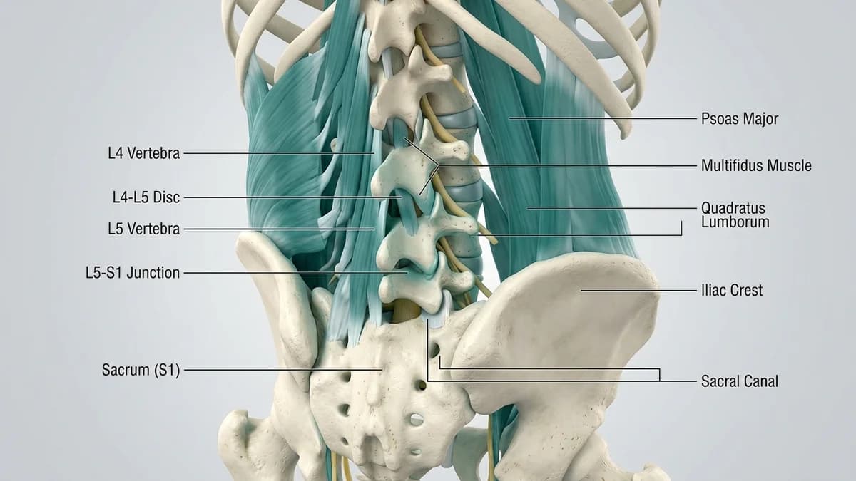
The anatomical mechanism underpinning the crossed SLR relies on the mechanical tethering of the thecal sac. When the unaffected leg is elevated, the ipsilateral L5 and S1 nerve roots are pulled taut and slide caudally through their respective neural foramina. Because these roots anchor directly into the central dural sheath, this downward traction pulls the entire thecal sac and the contralateral nerve roots caudally and medially across the midline. In a standard paracentral or shoulder herniation, where the disc lies lateral to the nerve root, this medial pull may actually decompress the nerve. However, when a herniation occupies an axillary position, nesting directly in the crook between the thecal sac and the emerging nerve root, the downward and medial movement forces the nerve root directly against the rigid disc fragment. This mechanical impingement reproduces acute radicular pain down the resting limb.
McKenzie Centralization, Neural Dynamic Sliders, and Graded Lumbar Mobilization
A positive well-leg raising test signals severe mechanical nerve root tethering, often indicating a large extrusion or sequestered fragment that requires careful handling. Traditional aggressive stretching or forceful spinal manipulation is strictly contraindicated. Physiotherapists prioritize gentle offloading and positional strategies, carefully evaluating whether prone lying with lateral pelvic shifting or gentle extension can begin to centralize the lesion. If an axillary herniation is present, pure extension may initially pinch the nerve root, requiring slight flexion or side-gliding adjustments to create space. Conservative rehabilitation focuses on quiet rest in the psoas position, gentle diaphragmatic breathing, and gradual introduction of non-provocative neural mobilization only after the crossed SLR sign has completely resolved.
Supported Psoas Unloading with Lateral Pelvic Shift Adjustment
Objective: Decompress the thecal sac and reduce mechanical impingement on axillary nerve roots
- 1Lie on your back on a firm, comfortable surface with a low pillow supporting your head and neck.
- 2Place an ottoman, supportive chair, or foam block under both lower legs so your hips and knees rest bent at 90 degrees.
- 3If resting flat provokes subtle leg pain, gently shift your pelvis 2 to 3 centimeters toward your pain-free side.
- 4Place a folded towel under your lumbar spine if mild support feels more comforting, ensuring no sharp pinching occurs.
- 5Rest quietly in this position for 10 minutes, breathing slowly through your nose to allow thecal sac tension to relax.
| Diagnostic Parameter | Standard Straight Leg Raise (SLR) | Crossed Straight Leg Raise (Fajersztajn Sign) |
|---|---|---|
| Limb Being Elevated | The symptomatic (painful) lower extremity is elevated passively by the examiner | The asymptomatic (well, pain-free) lower extremity is elevated passively by the examiner |
| Clinical Specificity and Sensitivity | High sensitivity (80-90%), but modest specificity (around 40-50%); frequent false positives | Exceptional specificity (88-97%), with lower sensitivity (around 25-40%); highly diagnostic |
| Underlying Patho-Anatomy | Indicates neural mechanosensitivity across any standard paracentral disc protrusion | Strongly indicates large disc extrusion, free sequestration, or an axillary disc location |
| Surgical Correlation Rate | Commonly positive in both non-operative and operative disc herniation presentations | Over 90% correlation with surgically confirmed structural disc herniations requiring close monitoring |
Because a positive crossed straight leg raise indicates a large disc extrusion or an axillary fragment that crosses near the midline, the risk of cauda equina compression is significantly higher. If you notice numbness around your groin, difficulty starting urination, or loss of bowel awareness, seek immediate emergency hospital care.
Frequently Asked Questions
Why does lifting my healthy leg make pain shoot down my bad leg?
The nerves for both legs connect to the same central spinal cord sac inside your spinal canal. When your physiotherapist lifts your healthy leg, it pulls that central sac downward and toward the midline. If you have a large disc herniation sitting in the inner crook of your nerve, this tug pulls the painful nerve straight against the disc, triggering pain in your opposite leg.
Do I need a doctor referral to treat a herniated disc on BookPhysio.in?
BookPhysio.in does not require a doctor referral. Under the National Commission for Allied and Healthcare Professions (NCAHP) Act 2021, patients across India enjoy direct access to autonomous registered physiotherapists. Through BookPhysio.in, you connect directly with verified clinicians and pay per session with transparent fees, completely free from advance package commitments, commission markups, or platform subscription charges.
Related recovery guides & articles
BookPhysio.in Editorial Team
The BookPhysio.in editorial team writes and maintains this content, checking it against reliable clinical sources. Content follows our medical review policy: general information only, and no page claims a clinical review until a named registered physiotherapist has signed it off.
Read our medical review policy