
Ankylosing Spondylitis Axial Extension Routine: Preventing Bamboo Spine Progression
In short
Ankylosing spondylitis drives progressive syndesmophyte bridging and rigid thoracic kyphosis. Learn evidence-based axial extension protocols, prone positioning routines, and domestic ergonomic adjustments to preserve upright spinal posture.
In this guide (6 sections)
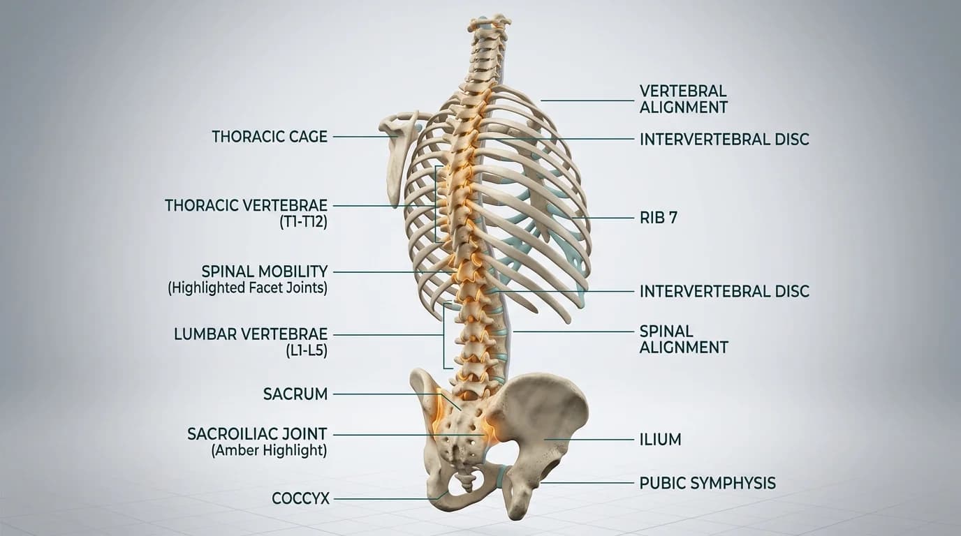
Ankylosing spondylitis is a chronic autoimmune inflammatory arthropathy that predominantly targets the axial skeleton, initiating at the sacroiliac joints and ascending through the lumbar, thoracic, and cervical spine. Chronic entheseal inflammation triggers repetitive micro-damage followed by aberrant repair cascades where fibrocartilage undergoes ossification, forming marginal syndesmophytes. Over decades, adjacent vertebral bodies fuse into a continuous, rigid osseous column colloquially termed bamboo spine. While modern biologic disease-modifying anti-rheumatic drugs suppress systemic inflammation, targeted axial physiotherapy is essential to ensure that newly formed bone consolidates in functional, upright extension rather than disabling forward kyphosis.
Domestic and Cultural Indian Realities
Managing ankylosing spondylitis in an Indian domestic setting presents unique postural challenges that must be systematically adapted. Traditional cross-legged floor sitting (sukhasana) during religious poojas and family meals forces the pelvis into posterior tilt, collapsing the lumbar lordosis and driving extreme thoracic flexion. During periods of active sacroiliitis, patients should sit on an elevated wooden pooja chowki measuring at least 18 inches in height, or use a firm dining chair with a supportive lumbar roll. Standard Indian squat commodes impose massive hip hyperflexion and pelvic torsion, which aggravates enthesitis at the ischial tuberosities; placing an over-toilet commode chair over the squat pan prevents biomechanical overload. In northern Indian winters, severe morning stiffness worsens due to falling ambient temperatures; patients benefit from taking an early morning warm water bucket bath before initiating bedside extension exercises, avoiding vigorous deep tissue massage with heated mustard oil which can trigger secondary acute synovitis.
Clinical Biomechanics and Inflammatory Pathology
The primary pathological site in axial spondyloarthritis is the enthesis, where tendons, ligaments, and the outer annulus fibrosus insert into vertebral cortical bone. In HLA-B27 positive individuals, mechanical tensile stress at these junctions stimulates local immune cells to release tumour necrosis factor alpha (TNF-alpha) and interleukin-17 (IL-17). This persistent cytokine cascade promotes osteoclastic bone erosion followed by bone morphogenetic protein activation, yielding chondrocyte proliferation and dense trabecular syndesmophytes. Because the anterior longitudinal ligament and zygapophyseal joints undergo progressive ankylosis, the spine loses sagittal flexibility. Without daily axial extension routines, gravity pulls the trunk forward into progressive hyperkyphosis, dramatically impairing horizontal forward gaze and restricting thoracic respiratory expansion.
Clinical Assessment and Functional Staging
Physiotherapy evaluation requires serial monitoring of objective anthropometric markers alongside patient-reported disease indices. The modified Schober test assesses lumbar flexion excursion, where an increase of less than 4 cm between skin markings placed 5 cm below and 10 cm above the lumbosacral junction indicates significant spinal restriction. Occiput-to-wall and tragus-to-wall distance measurements quantify early cervical and upper thoracic kyphotic deviation. Chest expansion measured circumferentially at the fourth intercostal space should exceed 2.5 cm; values below this threshold signal costovertebral joint ankylosis. Clinicians also record the Bath Ankylosing Spondylitis Disease Activity Index (BASDAI) and Functional Index (BASFI) to dose exercise intensity safely.
Targeted Mobility and Joint Preservation Exercises
Exercise prescription in axial spondyloarthritis focuses on maintaining spinal extension, strengthening deep back extensor musculature, and expanding thoracic cage volume. High-impact ballistic motions, heavy spinal loading, and repetitive trunk flexion exercises are contraindicated as they accelerate entheseal micro-damage. Instead, static positioning and controlled, unresisted active extension maintain sagittal balance.
Prone Sphinx Thoracic Extension and Scapular Retraction
Objective: Promote active thoracic extension and open the anterior chest wall without hyperextending the lumbar facet joints
- 1Lie prone on a firm treatment mat or firm wooden cot with your legs straight and toes relaxed.
- 2Prop your upper body on your forearms with elbows positioned directly beneath your shoulders and palms flat on the bed.
- 3Gently draw your shoulder blades backward and downward toward your back pockets while lengthening your neck.
- 4Breathe deeply into your lower ribs, holding the extended posture steadily for 20 seconds before gently lowering.
| Daily Activity | Conventional Habitual Pattern | Joint-Preserving Modification |
|---|---|---|
| Sleeping Surface | Sagging rope charpoy or thick coir mattress with multiple soft head pillows | Firm wooden cot with 12mm plywood base and single thin cervical contour roll |
| Spiritual Worship | Prolonged floor sitting cross-legged on a thin mat with rounded back | Elevated pooja chowki (18 inches) with firm back support maintaining upright spine |
| Desk Ergonomics | Slouched forward posture with low computer monitor forcing cervical flexion | Eye-level screen elevation with sternal chest lift and hourly standing back extensions |
| Sanitation Facility | Deep squatting over Indian ground pan provoking sacroiliac shearing | Raised Western toilet seat or sturdy over-commode support frame with bilateral rails |
Red Flags and Immediate Rheumatology Escalation
Immediate medical escalation is essential if a patient develops sudden red eye with deep pain and photophobia, which indicates acute anterior uveitis. Severe unremitting nocturnal thoracic pain accompanied by fever suggests spinal infection or vertebral discitis. Because fused ankylosed spines are brittle and act like long bones, minor falls or low-velocity vehicular bumps can cause chalk-stick fractures; any acute onset of localized spine tenderness or neurological deficits such as arm numbness, leg weakness, or bowel and bladder dysfunction requires immediate emergency hospital imaging.
Frequently Asked Questions
Can physiotherapy reverse bamboo spine once bony fusion has occurred?
Physiotherapy cannot dissolve or reverse mature ossified syndesmophytes once adjacent vertebrae have completely fused. However, targeted physiotherapy helps manage morning stiffness, maintain spinal mobility in unfused segments, and strengthen the paraspinal muscles. This preserves functional horizontal vision and expands thoracic breathing mechanics, preventing secondary postural decline.
How do I arrange a home physiotherapy visit for ankylosing spondylitis across Indian cities?
Under the National Commission for Allied and Healthcare Professions (NCAHP) Act 2021, patients in India can directly consult registered physiotherapists without a mandatory doctor referral. On BookPhysio.in, you connect with verified clinicians in your local city who visit your home. You pay the consultation fee directly to the physiotherapist at your appointment using cash or UPI, as BookPhysio.in does not collect booking fees online.
Related recovery guides & articles
BookPhysio.in Editorial Team
The BookPhysio.in editorial team writes and maintains this content, checking it against reliable clinical sources. Content follows our medical review policy: general information only, and no page claims a clinical review until a named registered physiotherapist has signed it off.
Read our medical review policy