
Anterior Cord Syndrome Management: Flexion Trauma, Motor Loss, and Rehab
In short
Anterior cord syndrome results in complete motor loss and analgesia with preserved deep proprioception, demanding specialised transfer and sensory compensatory protocols.
In this guide (6 sections)
Anterior cord syndrome is an incomplete spinal cord injury characterized by damage to the anterior two-thirds of the spinal cord, leaving the posterior columns intact. It is most commonly precipitated by severe acute hyperflexion cervical injuries (such as diving into shallow water or high-velocity vehicular crashes) causing retropulsed bone fragments or disc herniation, or by vascular insufficiency within the territory of the anterior spinal artery (such as during thoracic aortic aneurysm repairs). The clinical presentation features complete bilateral motor paralysis and loss of pain and temperature sensation below the lesion, with preserved light touch, vibration, and joint position sense.
Domestic and Cultural Indian Realities
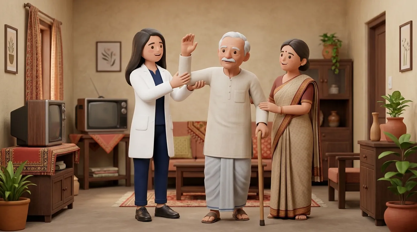
Coping with anterior cord syndrome in an Indian domestic setting presents unique clinical challenges. Because voluntary motor power is completely absent below the lesion, patients are fully dependent on wheelchairs for mobility and require sliding boards for bed and commode transfers. However, unlike individuals with complete transections (ASIA A), patients with anterior cord syndrome can clearly feel when their joints are positioned in flexion or extension and perceive the vibration of household machinery or vehicle engines. This preserved proprioceptive awareness provides a valuable neurological foundation for learning seated posture and transfer mechanics, although the complete lack of protective pain sensation exposes the skin to severe friction and burn risks.
Clinical Biomechanics and Neurological Recovery
The anterior spinal artery supplies the anterior two-thirds of the spinal cord, encompassing the anterior horns (motor neurons), the lateral corticospinal tracts (voluntary movement), and the spinothalamic tracts (pain and temperature). Infarction or contusion within this vascular bed results in extensive bilateral necrosis. However, the posterior spinal arteries, which supply the dorsal columns (fasciculus gracilis and cuneatus), maintain adequate perfusion. Biomechanically, because dorsal column inputs remain intact, the brain receives accurate, continuous kinesthetic feedback regarding pelvic tilt and joint angles, enabling patients to maintain seated balance using visual and somatosensory cues.
Functional Movement Analysis and Clinical Assessment
Neurological classification requires careful sensory examination: applying a 128 Hz tuning fork to the medial malleolus and anterior superior iliac spine demonstrates intact vibration sense, and toe position testing confirms preserved proprioception. In contrast, pinprick testing reveals complete analgesia below the lesion level, and motor examination confirms grade 0/5 power in all sub-lesional key muscles. Upper extremity strength must be evaluated thoroughly: if the lesion is situated in the mid-to-lower cervical spine, the patient functional potential mirrors that of complete tetraplegia or paraplegia.
Targeted Rehabilitation and Functional Independence Protocol
Rehabilitation focuses on capitalizing on preserved kinesthetic awareness while establishing rigorous compensatory strategies for absent pain sensation. Transfer training utilizes head-hips mechanics: the patient can accurately perceive when the pelvis is properly aligned on a sliding board, speeding motor learning. A strict 15-minute pressure relief schedule is mandated, because although the patient can feel the position of their buttocks on the cushion, they cannot feel ischemic pain or tissue starvation. Environmental modifications include thermostatic controls on bathroom water geysers and protective clothing over insensate limbs.
Proprioceptive-Guided Seated Weight Shift and Postural Centring
Objective: Utilize preserved dorsal column sensation to maintain optimal pelvic alignment in seating
- 1Sit upright in your wheelchair with your feet flat on the footrests and brakes locked.
- 2Place your palms flat on your thighs or on the wheelchair armrests.
- 3Close your eyes and focus on the feeling of weight distribution beneath both ischial tuberosities.
- 4Slowly shift your torso 2 inches to the right, feeling the increased pressure and hip joint compression.
- 5Smoothly return to the exact centre, verifying that weight feels equally shared between both buttocks.
- 6Lean forward 30 degrees, feeling the weight transition to the anterior thighs, then return to neutral.
| Spinal Cord Tract | Anatomical Location and Status | Clinical Functional Manifestation |
|---|---|---|
| Lateral Corticospinal Tract | Anterior two-thirds; completely disrupted | Bilateral motor paralysis below the lesion level |
| Lateral Spinothalamic Tract | Anterior two-thirds; completely disrupted | Total loss of pain and temperature sensation below lesion |
| Posterior Dorsal Columns | Posterior one-third; intact and well-perfused | Preserved joint position sense, vibration, and deep touch |
| Autonomic Pathways | Descending sympathetic tracts disrupted if T6 or above | Neurogenic bowel, neurogenic bladder, risk of autonomic dysreflexia |
Red Flags and Emergency Escalation
Because pain sensation is entirely absent below the lesion, patients with anterior cord syndrome will not feel burns from hot bathing water, cuts from sharp furniture edges, or the pain of an acute bone fracture. Daily visual skin inspection from head to toe is essential. For injuries at T6 or higher, any unrecognized skin damage or urinary blockage can trigger a sudden hypertensive crisis through autonomic dysreflexia. In acute cases following trauma, watch for ascending motor weakness or respiratory distress, which signals expanding cord edema requiring emergency surgical decompression.
Frequently Asked Questions
Does feeling deep touch mean that motor power will eventually return?
Not necessarily. The sensory signals you feel for touch and vibration travel along the back of the spinal cord (dorsal columns), which survived the injury. The motor nerve fibers that control muscle movement travel in the front of the cord, which suffered severe damage. While some motor recovery is possible, recovery depends on the extent of sparing in the anterior corticospinal pathways.
How do I arrange specialised home physiotherapy for anterior cord syndrome?
Under the NCAHP Act 2021, registered physiotherapists in India can be accessed directly for domiciliary neurological care. On BookPhysio.in, book an experienced physiotherapist who will structure a customized transfer, seating, and pressure care program at home. Consultation fees are paid directly to the attending clinician at your appointment.
Related recovery guides & articles
BookPhysio.in Editorial Team
The BookPhysio.in editorial team writes and maintains this content, checking it against reliable clinical sources. Content follows our medical review policy: general information only, and no page claims a clinical review until a named registered physiotherapist has signed it off.
Read our medical review policy