
Cauda Equina Syndrome vs Conus Medullaris: Decompression and Neuro-Rehab
In short
Differentiating cauda equina syndrome from conus medullaris lesions dictates surgical urgency and shapes lower motor neuron neuro-rehabilitation protocols.
In this guide (6 sections)
Compressive pathologies of the lumbosacral junction present along a clinical continuum between conus medullaris syndrome and cauda equina syndrome. The conus medullaris represents the tapered terminal end of the true spinal cord, terminating between the lower border of the L1 and upper L2 vertebrae in adults. In contrast, the cauda equina is an intradural collection of peripheral lumbosacral nerve roots (L2 through S5) descending through the lumbar cistern. While conus lesions produce a mixed upper and lower motor neuron picture with early bilateral sphincter paralysis, cauda equina compression represents a pure lower motor neuron disorder characterized by severe asymmetric radicular pain, flaccid paraparesis, saddle anesthesia, and areflexic bladder dysfunction.
Domestic and Cultural Indian Realities
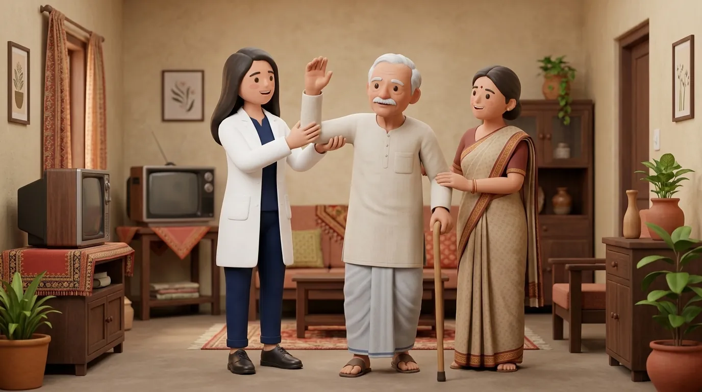
In Indian clinical practice, cauda equina syndrome is frequently misdiagnosed in its initial stages as benign muscular low back pain or simple sciatica. Patients often endure weeks of rest, heating pads, or local unscientific spine manipulations while progressive bladder retention develops silently. By the time painless overflow urinary incontinence and complete perineal saddle anesthesia manifest, irreversible nerve root ischemia has occurred. Following emergency surgical decompression, domiciliary rehabilitation must address home toileting, clean intermittent self-catheterization on domestic beds, and custom orthotic prescription for bilateral foot drop to restore community ambulation.
Clinical Biomechanics and Neurological Recovery
Because the nerve roots of the cauda equina belong to the peripheral nervous system, they possess Schwann cell sheaths capable of slow axonal regeneration (at approximately 1 mm per day), offering greater long-term recovery potential than the central cord tracts of the conus medullaris. However, prolonged mechanical compression by a massive extruded lumbar disc or fracture fragment induces endoneurial edema, capillary thrombosis, and Wallerian degeneration. Biomechanically, denervation of the tibialis anterior, gastrocnemius, and gluteus medius disrupts the kinetic gait chain, producing a high-stepping steppage gait or severe Trendelenburg lurch that requires external orthotic stabilization.
Functional Movement Analysis and Clinical Assessment
Clinical clinical examination mandates meticulous dermatomal and myotomal charting. Saddle sensation across dermatomes S2 through S5 (perineum, perianal region, and inner thighs) is tested with cotton wool and pinprick. The bulbocavernosus reflex (elicited by squeezing the glans penis or clitoris and observing anal sphincter contraction) is preserved in conus lesions but absent in cauda equina syndrome. Motor power in key lumbosacral myotomes, hip flexors (L2), knee extensors (L3), ankle dorsiflexors (L4), long toe extensors (L5), and ankle plantarflexors (S1), is graded, and post-void residual bladder volume is measured via bedside ultrasound.
Targeted Rehabilitation and Functional Independence Protocol
Post-operative rehabilitation follows a progressive neuro-muscular re-education protocol. In Phase 1, gentle neural mobilization (flossing) of the sciatic and femoral nerves is introduced to prevent epidural fibrosis and root tethering. In Phase 2, patients learn pelvic floor muscle re-education combined with clean intermittent self-catheterization (CIC) to prevent bladder overdistension. In Phase 3, gait training incorporates custom lightweight carbon-fiber or polypropylene ankle-foot orthoses (AFOs) to stabilize foot drop, progressing from walking frames to independent community ambulation.
Seated Sciatic Nerve Slider and Neural Gliding Drill
Objective: Promote gentle neural mobility and reduce perineural adhesions following decompression
- 1Sit upright on a firm, comfortable chair with your thighs fully supported and feet flat on the floor.
- 2Slowly extend your affected knee straight out in front while simultaneously tilting your head backward to look at the ceiling.
- 3Pause for 1 second at the top of the motion without forcing through sharp nerve pain.
- 4Simultaneously bend your knee back down while tucking your chin toward your chest.
- 5Maintain a smooth, continuous, rhythmic cycling movement between the two positions.
- 6Ensure the movement remains completely pain-free, acting as a gentle neural flossing motion.
| Clinical Feature | Conus Medullaris Syndrome | Cauda Equina Syndrome |
|---|---|---|
| Anatomical Lesion Level | L1 to L2 vertebral level (Terminal spinal cord) | Below L2 vertebral level (Lumbosacral nerve roots) |
| Motor Deficit Symmetry | Bilateral, symmetric; mixed UMN and LMN signs | Markedly asymmetric, patchy; pure LMN flaccidity |
| Radicular Pain Presentation | Mild, bilateral low back pain; minimal radicular pain | Severe, sharp, radiating sciatica in legs and perineum |
| Bulbocavernosus Reflex | Preserved / Hyperactive (Sacral reflex arc intact) | Absent (Afferent and efferent roots severed) |
Red Flags and Emergency Escalation
Cauda equina compression is a true surgical emergency. If any individual experiencing back pain or sciatica develops acute urinary retention, inability to feel toilet paper when wiping (saddle anesthesia), progressive bilateral leg weakness, or fecal incontinence, they must be transported immediately to an emergency department for urgent MRI and neurosurgical decompression within 24 to 48 hours. Delayed decompression beyond 48 hours is strongly associated with permanent bladder paralysis, irreversible sexual dysfunction, and chronic neuropathic pain.
Frequently Asked Questions
Why is emergency surgery required so quickly for cauda equina syndrome?
The nerve roots that control bladder, bowel, and sexual function are delicate and vulnerable to pressure. If mechanical compression cuts off their blood supply for more than 48 hours, the nerve cells suffer irreversible ischemic death, leading to lifelong bladder paralysis and loss of pelvic sensation even if surgery is performed later.
How do I arrange home physiotherapy for foot drop and bladder re-education after surgery?
Under the NCAHP Act 2021, registered physiotherapists provide direct rehabilitation without requiring doctor referrals. On BookPhysio.in, you can book an experienced neuro-physiotherapist who will fit orthoses, train nerve gliding, and structure pelvic floor exercises in your home. Fees are paid directly to the clinician at your appointment.
Related recovery guides & articles
BookPhysio.in Editorial Team
The BookPhysio.in editorial team writes and maintains this content, checking it against reliable clinical sources. Content follows our medical review policy: general information only, and no page claims a clinical review until a named registered physiotherapist has signed it off.
Read our medical review policy