
Chronic Charcot Foot Management: Rocker-Bottom Soles and Custom Orthotics
In short
When acute Charcot neuroarthropathy resolves, it frequently leaves a consolidated rocker-bottom foot deformity with high focal midfoot pressures. Discover how custom orthotics and rocker soles restore mobility.
In this guide (6 sections)
Chronic Charcot neuroarthropathy (Eichenholtz Stage 3) represents the permanent consolidation phase following acute destructive inflammation. In many cases, midfoot collapse results in a classical rocker-bottom deformity, where the cuboid, navicular, or medial cuneiform bones protrude downward, forming the most inferior weight-bearing point of the foot. Without the natural arch structure, standard footwear concentrates tremendous peak vertical pressures and shear stresses directly onto this bony midfoot prominence. Clinical rehabilitation focuses on lifelong pressure redistribution using custom total contact orthoses, rigid rocker-bottom footwear, and gait retraining to shield the compromised midfoot from ulceration and osteomyelitis.
Domestic and Cultural Indian Realities
Managing a chronic rocker-bottom Charcot foot in an Indian home requires permanent abandonment of all conventional sandals, chappals, and barefoot walking. Because the midfoot convexity bears the brunt of every step, walking barefoot across hard tile or stone floors will ulcerate the skin within days. In many households, patients struggle with traditional social expectations during religious ceremonies or family meals where shoes are frowned upon indoors. Families must accept that medical footwear must be worn inside the living areas. A dedicated clean pair of indoor Charcot shoes with custom moulded footbeds and stiff rocker soles should be designated exclusively for domestic use. When entering sacred pooja rooms, using clean washable shoe covers over prescription orthopaedic shoes maintains spiritual sanctity while preserving tissue integrity.
Clinical Biomechanics and Neuropathic Breakdown
In a normal foot, ground reaction forces during the stance phase of gait pass smoothly from the calcaneus through the lateral longitudinal arch to the metatarsal heads, with the midfoot remaining elevated. In a consolidated rocker-bottom Charcot foot, the apex of the collapsed midfoot becomes the primary weight-bearing pillar. Peak plantar pressures over this bony apex regularly exceed 800 kilopascals, far above the capillary perfusion threshold. Furthermore, the shortened Achilles tendon creates an equinus contracture that accelerates midfoot loading during terminal stance. A rigid rocker sole addresses this pathomechanics by incorporating a curvature with an apex positioned just proximal to the midfoot prominence, allowing the patient to roll through the step without bending the shoe or concentrating pressure over the collapsed tarsal bones.
Clinical Assessment and Neuropathy Screening
Assessment involves precise physical evaluation of foot morphology, identifying the location and extent of midfoot bony prominences, joint rigidity, and skin thickness over the apex. Dynamic baropodometric pressure mapping is utilized to quantify exact peak pressures in standing and walking. The clinician verifies that the Charcot process is quiescent by confirming normal skin temperature symmetry (within 1 degree Celsius) and checking for the absence of localized oedema. Ankle dorsiflexion is measured with the knee straight to evaluate for gastrocnemius-soleus tightness, and the skin is examined for pre-ulcerative calluses, redness, or subcutaneous seroma formation.
Targeted Offloading and Intrinsic Foot Conditioning
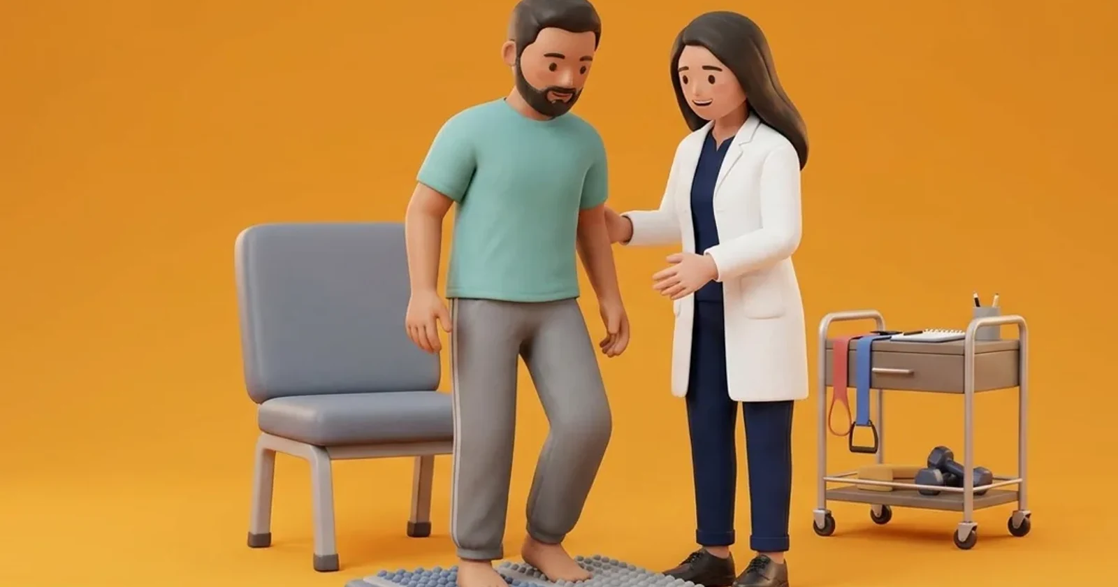
Long-term management relies on custom-moulded total contact orthoses manufactured using multi-density materials (such as Plastazote combined with high-density EVA) with deep relief recesses carved directly beneath the midfoot prominence. Footwear must incorporate a stiff carbon fibre or composite shank and a rigid external rocker sole. Physiotherapy emphasizes gait retraining to shorten stride length, reduce walking speed, and ensure smooth heel-to-toe rollover without sudden ground impacts. Seated non-weight bearing calf stretches help alleviate equinus forces that drive the midfoot downward.
Seated Long-Sitting Calf Towel Stretch
Objective: Lengthen tight posterior calf structures to reduce mechanical midfoot loading
- 1Sit on a firm bed or exercise mat with legs extended straight in front of you.
- 2Loop a non-elastic cotton towel or strap around the ball of the foot, holding the ends in both hands.
- 3Keeping your knee completely straight, gently pull the towel toward your torso until you feel a comfortable stretch in your calf.
- 4Maintain the stretch steadily for 20 to 30 seconds without bouncing.
- 5Gently release the tension, rest for 10 seconds, and repeat on the same leg.
| Biomechanical Component | Engineering Specification | Clinical Mechanism |
|---|---|---|
| Custom Total Contact Insole | Multi-density Plastazote top layer with EVA base and carved midfoot well | Distributes plantar load across the entire foot surface, eliminating focal midfoot peak pressure |
| Rigid Rocker-Bottom Outer Sole | Apex positioned 55-60% of shoe length from heel, 25-degree toe spring | Replaces sagittal plane foot motion, eliminating midfoot bending and forefoot propulsive shear |
| High-Top Ankle Collar / Boot | Rigid medial and lateral thermoplastic counters extending above malleoli | Restricts subtalar eversion and ankle inversion, stabilizing coronal plane alignment |
| Extra-Depth Broad Toe Box | 12mm added vertical depth with smooth soft leather lining | Accommodates dorsal claw toe deformities without frictional blister formation |
Red Flags and Urgent Diabetic Foot Escalation
Immediate medical review is required if the patient notices localized warmth, swelling, or redness returning over the midfoot, which may signal reactivation of acute Charcot arthropathy. The appearance of a dark blood blister, skin abrasion, or open breakdown over the rocker-bottom apex represents an emergency requiring total non-weight bearing until evaluated by a podiatric team. Any systemic fever or purulent drainage demands hospital admission to exclude deep bone infection.
Frequently Asked Questions
Can surgery fix the rocker-bottom bone bump instead of wearing specialised shoes?
Surgical exostectomy (shaving down the bony bump) or midfoot reconstruction with intramedullary beaming plates may be considered if recurrent ulcers develop despite optimal orthotic footwear. However, Charcot bone is often fragile and surgical complication rates are high; conservative management with custom footwear remains the preferred first-line strategy.
How often should custom Charcot footwear and insoles be replaced?
Custom orthotic insoles compress under repeated body weight, losing their protective cushioning over time. Insoles should be evaluated by a physiotherapist every 4 to 6 months and typically replaced every 6 to 12 months. The outer rocker sole and shoe integrity must also be checked regularly for uneven wear patterns that could alter walking mechanics.
Related recovery guides & articles
BookPhysio.in Editorial Team
The BookPhysio.in editorial team writes and maintains this content, checking it against reliable clinical sources. Content follows our medical review policy: general information only, and no page claims a clinical review until a named registered physiotherapist has signed it off.
Read our medical review policy