
Custom Moulded Total Contact Insoles for High-Risk Diabetic Foot Offloading
In short
Custom-moulded total contact insoles dramatically reduce peak plantar pressures by increasing contact surface area across the foot. Discover clinical fabrication and offloading protocols.
In this guide (6 sections)
Custom-moulded total contact insoles (TCIs) are prescribed orthotic devices designed to prevent neuropathic ulceration in high-risk diabetic feet. The fundamental biomechanical principle underlying total contact orthoses is derived from the physical definition of pressure: Pressure equals Force divided by Area. In an unsupported neuropathic foot, walking forces are concentrated onto tiny, prominent anatomical areas (such as the first and fifth metatarsal heads and calcaneus), resulting in destructive focal peak pressures. A custom TCI is sculpted to capture the exact anatomical contours of the patient plantar arches, expanding the contact surface area to the entire footprint. This intimate contouring redistributes forces away from vulnerable bony prominences onto forgiving soft tissues, reducing peak focal pressures by up to 40 percent.
Domestic and Cultural Indian Realities
In Indian clinical settings, many diabetic patients invest in off-the-shelf gel or foam insoles purchased online, believing they provide adequate medical protection. However, generic flat insoles fail to provide total contact contouring and quickly bottom out under body weight, offering zero pressure relief for rigid deformities or fallen arches. Furthermore, patients often transfer insoles into standard footwear that lacks sufficient vertical depth, creating severe dorsal shoe friction that ulcerates the tops of the toes. Custom insoles must be fabricated from 3D volumetric plaster casts, foam impression boxes, or optical scans taken in subtalar neutral, and fitted exclusively into extra-depth prescription footwear.
Clinical Biomechanics and Neuropathic Breakdown
Effective diabetic insole design relies on a multi-density sandwich construction. The top interface layer must consist of a closed-cell, heat-mouldable polyethylene foam (such as Plastazote), which possesses low shear resistance and dynamically moulds to subtle foot contours without causing skin friction. Beneath this interface sits a resilient mid-layer of microcellular rubber (MCR) or Poron, which absorbs vertical impact and recovers its shape after compression. The base layer is made of firm high-density ethylene-vinyl acetate (EVA) or cork to support the medial longitudinal arch. Carved relief wells, metatarsal pads, or horseshoe cutouts are incorporated directly beneath prominent metatarsal heads or past ulcer sites to eliminate localized pressure spikes entirely.
Clinical Assessment and Neuropathy Screening
Assessment begins with non-weight bearing semi-weight bearing physical examination of foot morphology, evaluating arch height, joint flexibility, and digital alignment. The clinician identifies previous ulcer sites, hyperkeratotic calluses, and bony prominences. Dynamic in-shoe baropodometric pressure measurement or Harris mat footprinting confirms exact pressure distribution. Sensory mapping with 10g monofilaments determines the boundaries of loss of protective sensation, while pedal pulses verify adequate blood flow.
Targeted Offloading and Intrinsic Foot Conditioning
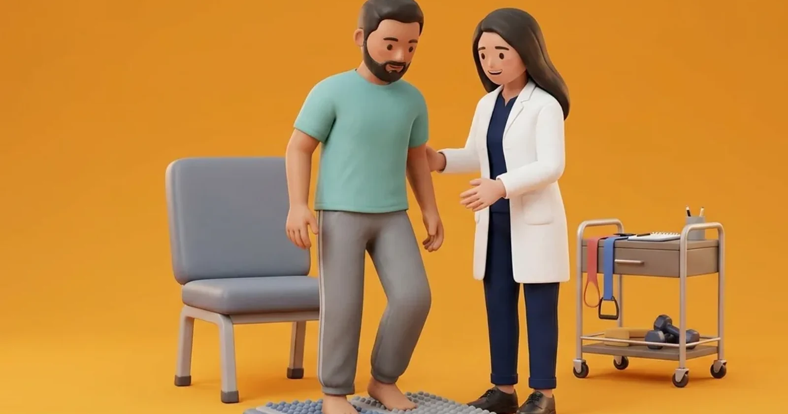
Once custom insoles are fitted, the physiotherapist evaluates the patient gait to ensure smooth transition from heel strike to toe roll. Patients are instructed to inspect their insoles weekly for signs of localized compression, cracking, or bottoming out. Because Plastazote compresses permanently over months of use, insoles must be evaluated every 4 to 6 months and refurbished or replaced when cushioning thickness degrades.
Seated Arch Awareness and Insole Alignment Drill
Objective: Familiarize the patient with custom insole contours without full body weight impact
- 1Sit comfortably in a sturdy chair with your custom insoles placed inside your prescription shoes.
- 2Place both feet flat on the floor with knees bent at 90 degrees.
- 3Gently press your arch downward into the contoured arch support of the insole for 5 seconds.
- 4Relax the downward pressure and gently rock weight toward your heels for 5 seconds.
- 5Rock weight smoothly toward the balls of your feet without lifting your heels.
- 6Repeat the gentle pressure distribution cycle smoothly.
| Insole Layer / Component | Material & Durometer Specification | Clinical Biomechanical Function |
|---|---|---|
| Top Interface Layer | Plastazote (Pink/White), Shore A 15-20, thickness 3-5mm | Direct skin contact; conforms to foot; minimizes friction and shear |
| Mid Cushioning Layer | Poron / Medical MCR, Shore A 20-25, thickness 3-5mm | Absorbs dynamic vertical shock; high resilience against bottoming out |
| Base Structural Layer | High-density EVA / Subortholen, Shore A 45-55, thickness 5-8mm | Maintains medial arch contour; prevents insole twisting in shoe |
| Metatarsal Offloading Dome | Soft Poron pad placed proximal to metatarsal heads 2-4 | Transfers load to metatarsal shafts, offloading prominent heads |
Red Flags and Urgent Diabetic Foot Escalation
Immediate medical escalation is necessary if removing shoes and insoles reveals localized redness, blistering, or bleeding over the arch or metatarsal pads. An insole that exerts excessive upward pressure under a stiff, arthritic arch can cause pressure ulceration along the medial navicular bone. The patient must stop wearing the insole immediately and return to the orthotist for insole adjustment.
Frequently Asked Questions
Can I move my custom diabetic insole into my regular dress shoes or sneakers?
Usually not. Custom total contact insoles are substantially thicker (approximately 10 to 14 millimetres total) than factory insoles. Placing them inside standard shoes severely reduces the internal volume, squeezing your toes against the shoe upper and causing blisters. They should only be used inside extra-depth diabetic shoes designed to accommodate them.
How often do custom total contact insoles need to be replaced?
The top Plastazote foam layer gradually packs down and loses its cushioning properties under body weight. For active individuals, insoles should be inspected by a physiotherapist every 4 to 6 months and typically replaced or resurfaced every 9 to 12 months to maintain effective offloading.
Related recovery guides & articles
BookPhysio.in Editorial Team
The BookPhysio.in editorial team writes and maintains this content, checking it against reliable clinical sources. Content follows our medical review policy: general information only, and no page claims a clinical review until a named registered physiotherapist has signed it off.
Read our medical review policy