
Diabetic Amyotrophy Rehabilitation: Femoral Nerve Neuropathy and Pelvic Strengthening
In short
Diabetic amyotrophy causes sudden, severe proximal thigh wasting and debilitating weakness in hip and knee musculature. Discover structured pelvic strengthening and gait rehabilitation protocols.
In this guide (6 sections)
Diabetic amyotrophy, also known clinically as diabetic lumbosacral radiculoplexus neuropathy (DLRPN) or Bruns-Garland syndrome, is an uncommon and debilitating manifestation of diabetes. Unlike symmetrical distal polyneuropathy, diabetic amyotrophy typically presents acutely or subacutely with severe unilateral neuropathic pain in the lumbar spine, hip, anterior thigh, or groin, followed rapidly by profound muscle wasting and motor weakness of the quadriceps, iliopsoas, and hip adductors. Resulting from microvascular ischaemia and immune-mediated epineurial microvasculitis, this proximal motor deficit impairs sit-to-stand transfers and causes frequent knee buckling during walking. Physiotherapy rehabilitation focuses on targeted pelvic stabilization, progressive closed-chain quadriceps strengthening, and assistive device prescription to prevent falls and restore functional independence.
Domestic and Cultural Indian Realities
In Indian domestic environments, diabetic amyotrophy causes severe functional disruption because daily life heavily relies on lower-limb proximal strength. Rising from low wooden cots, rope charpoys, or traditional floor seating becomes impossible without pulling on nearby furniture. Using traditional Indian squat commodes is exceptionally dangerous; when a patient quadriceps buckle under body weight, catastrophic falls, hip fractures, or knee dislocations occur. Families must immediately install a raised toilet seat or bedside commode with sturdy bilateral armrests. Low beds must be elevated with hardwood risers so the mattress sits at knee level (approximately 20 inches from the floor). Walking frames with front wheels are essential to support the patient during indoor mobility, preventing sudden knee collapse on smooth tiled floors.
Clinical Biomechanics and Neuropathic Breakdown
The pathophysiology of diabetic amyotrophy involves an immune-mediated inflammatory microvasculitis targeting the lumbosacral plexus and proximal femoral nerve branches (L2, L3, L4). Microvascular occlusion produces ischaemic infarction of nerve bundles, causing severe denervation of the rectus femoris, vastus lateralis, vastus medialis, and psoas major. Biomechanically, loss of quadriceps eccentric control eliminates the knee extensor moment required during the loading response of the gait cycle. To avoid knee collapse, patients compensate by leaning their trunk forward (compensatory hip flexion) or locking the knee into hyperextension (genu recurvatum) during stance, which stretches the posterior knee capsule and leads to progressive joint instability.
Clinical Assessment and Neuropathy Screening
Assessment begins with detailed manual muscle testing (MMT) of the hip flexors, adductors, abductors, and knee extensors, graded on the Medical Research Council (MRC) scale. Quadriceps circumference is measured bilaterally at 10 and 15 centimetres proximal to the superior patellar pole to document the degree of atrophy. Deep tendon reflexes demonstrate a markedly diminished or absent patellar tendon reflex with a preserved Achilles reflex. Electromyography and nerve conduction studies confirm multifocal denervation in lumbosacral paraspinal and femoral nerve distributions, distinguishing amyotrophy from compressive lumbar disc herniation, spinal stenosis, or inflammatory polymyositis.
Targeted Offloading and Intrinsic Foot Conditioning
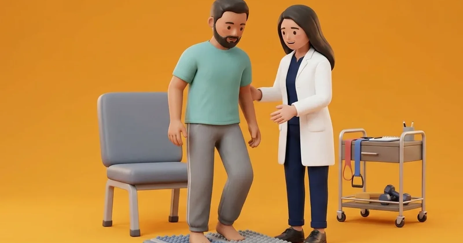
Rehabilitation progresses carefully from gravity-eliminated active-assisted exercise to closed-chain functional strengthening as re-innervation occurs over months. Initial training emphasizes pelvic bridging, isometric quadriceps sets, and straight leg raises in supine. As motor recruitment improves, patients practice seated sit-to-stand drills from elevated chair heights using arm support. To protect the knee from sudden hyperextension or buckling, a lightweight knee-ankle-foot orthosis (KAFO) or a hinged knee brace with extension stop may be prescribed for walking.
Supine Isometric Pelvic Bridging with Thigh Pillow Squeeze
Objective: Strengthen gluteus maximus, hamstrings, and hip adductors without joint shear
- 1Lie flat on your back on a firm bed with knees bent and feet resting flat on the mattress.
- 2Place a firm cushion or folded towel between your knees.
- 3Gently squeeze the cushion between your knees while drawing your navel in toward your spine.
- 4Press downward through your heels and lift your hips upward into a straight line from shoulders to knees.
- 5Hold the elevated bridge steadily for 5 seconds while breathing calmly.
- 6Slowly lower your hips back to the bed and relax for 3 seconds.
| Clinical Phase | Symptoms & Neuromuscular Status | Physiotherapy & Mobility Protocol |
|---|---|---|
| Acute Pain Phase (Weeks 0 to 6) | Severe burning thigh/groin pain; sleep disruption; early weakness | Pain management positioning; gentle passive ROM; wheeled walker for transfers |
| Atrophic Wasting Phase (Weeks 6 to 16) | Stabilization of pain; marked quadriceps wasting; knee buckling | Isometric sets, supine pelvic bridging, sit-to-stand from raised chair |
| Early Recovery Phase (Months 4 to 12) | Slow re-innervation; gradual motor improvement (MRC grade 3 to 4) | Closed-chain mini-squats, stationary recumbent cycling, elbow crutches |
| Functional Consolidation (Year 1 to 2) | Residual mild weakness or full recovery; endurance deficits | Stair training, balance retraining, long-term walking programme |
Red Flags and Urgent Diabetic Foot Escalation
Immediate medical escalation is necessary if a patient experiences sudden loss of bowel or bladder control, numbness in the saddle area (perineum), or rapidly progressive bilateral lower limb paralysis, which signals cauda equina syndrome requiring emergency neurosurgical decompression. If the patient sustains an unobserved fall with inability to bear weight, urgent radiographs are required to exclude femoral neck fractures.
Frequently Asked Questions
Does muscle strength return after diabetic amyotrophy?
Yes, but recovery is gradual. Diabetic amyotrophy has a positive long-term prognosis, with nerve regeneration and muscle recovery occurring slowly over 12 to 24 months. Although some residual asymmetry or mild quadriceps weakness may persist, structured physiotherapy and tight glycaemic control allow the vast majority of patients to regain independent walking.
How does diabetic amyotrophy differ from ordinary diabetic neuropathy?
Ordinary diabetic polyneuropathy develops slowly over years, affects both feet equally, causes numbness and tingling starting in the toes, and rarely causes severe proximal muscle wasting. In contrast, diabetic amyotrophy starts suddenly, often on one side, causes intense thigh pain, and selectively weakens the large thigh and hip muscles responsible for standing.
Related recovery guides & articles
BookPhysio.in Editorial Team
The BookPhysio.in editorial team writes and maintains this content, checking it against reliable clinical sources. Content follows our medical review policy: general information only, and no page claims a clinical review until a named registered physiotherapist has signed it off.
Read our medical review policy