
Juvenile Dermatomyositis: Muscle Weakness and Corticosteroid Recovery
In short
Managing Juvenile Dermatomyositis requires balancing active inflammatory containment with staged, fatigue-monitored physical rehabilitation.
In this guide (6 sections)
Juvenile Dermatomyositis (JDM) is the most common paediatric inflammatory idiopathic myopathy, characterised by systemic autoimmune microangiopathy affecting skeletal muscle tissue and skin. Children typically present between five and ten years of age with pathognomonic cutaneous signs (heliotrope violaceous rash around the eyelids and Gottron papules over finger joints) combined with progressive, symmetrical proximal muscle weakness affecting the pelvic and shoulder girdles, neck flexors, and trunk. While modern medical therapy using high-dose corticosteroids, methotrexate, and intravenous immunoglobulin (IVIG) halts acute immune-mediated vascular necrosis, prolonged corticosteroid use introduces severe secondary complications: steroid-induced myopathy, osteopenia, rapid weight gain, and muscle atrophy. Paediatric physiotherapy is essential throughout the disease trajectory, carefully progressing from gentle passive maintenance during active vasculitis toward progressive resistive strengthening and contracture management during medical remission.
Domestic and Cultural Indian Realities
In India, initial diagnosis of JDM is frequently delayed for months, as facial rashes are often misattributed to sunburn, fungal ringworm, or cosmetic allergies, while rising fatigue and difficulty climbing stairs are mistaken for growing pains or nutritional anaemia. Furthermore, the extreme sun sensitivity (photosensitivity) inherent to JDM presents massive daily challenges under the harsh tropical Indian sun. Travelling to school, walking across open school courtyards, and participating in outdoor physical education triggers massive cutaneous and systemic flare-ups. Paediatric physiotherapists must coordinate with schools to ensure strict sun protection: tinted classroom windows, UV-protective clothing, broad-spectrum sunscreen application every two hours, and holding all physiotherapy sessions indoors in cool, climate-controlled rooms.
Clinical Biomechanics and Neurological Development
The pathobiology of JDM involves complement-mediated (C5b-9 membrane attack complex) lysis of endomysial capillary endothelial cells, leading to microvascular occlusion, focal muscle ischaemia, and perifascicular myofibre atrophy. Biomechanically, profound weakness of the gluteus maximus, gluteus medius, and quadriceps severely impairs the child ability to rise from a low chair or climb stairs without pushing hands against knees (Gowers sign). Furthermore, ischaemic muscle injury triggers intense fibroblastic proliferation, causing rapid, severe flexion contractures at the elbows, hips, knees, and ankles. In longstanding under-treated cases, dystrophic calcification (calcinosis cutis and calcinosis universalis) deposits rigid calcium phosphate crystals in subcutaneous sheets, mechanically locking joint excursions.
Clinical Assessment and Developmental Staging
Clinical evaluation follows the International Myositis Assessment and Clinical Studies (IMACS) criteria. Muscle strength is objectively quantified using the Childhood Myositis Assessment Scale (CMAS, scoring 14 functional motor tasks out of 52) and the Manual Muscle Testing-8 (MMT-8, grading 8 proximal and distal muscle groups out of 80). Laboratory monitoring tracks serum muscle enzymes: creatine kinase (CK), lactate dehydrogenase (LDH), and alanine aminotransferase (ALT). Passive joint goniometry monitors contractures, and skin inspection screens for emerging calcinosis nodules.
Targeted Rehabilitation and Mobility Exercises
Historically, exercise during active myositis was avoided out of fear of worsening inflammation. Contemporary clinical trials confirm that carefully monitored, non-fatiguing active exercise is safe and anti-inflammatory, promoting muscle revascularization. In the acute phase, therapy emphasizes gentle passive range of motion and positioning. As medical therapy controls acute inflammation (normalizing CK levels), treatment advances to isometric contractions, closed-chain functional strengthening, and submaximal aerobic conditioning.
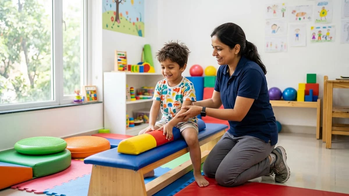
Closed-Chain Seated Bench Sit-to-Stand and Pelvic Alignment
Objective: Strengthen proximal gluteal and quadriceps musculature without eccentric muscle damage
- 1Seat the child upright on a firm, height-adjustable therapy bench with feet flat on the floor.
- 2Ensure the bench height is set so the child hips and knees are at a comfortable 90-degree angle.
- 3Instruct the child to cross their arms over their chest to prevent pushing on their thighs.
- 4Guide the child to lean forward from the hips (nose over toes) and press down through both heels to stand tall.
- 5Hold the upright standing posture steadily for 3 seconds, squeezing the gluteal muscles.
- 6Slowly and smoothly lower back down to the seated bench in a controlled manner, avoiding plumping down.
| Disease Stage | Clinical Characteristics & Muscle Enzymes | Physiotherapy Management Strategy |
|---|---|---|
| Acute Inflammatory Phase | Marked muscle weakness (CMAS <25), elevated CK/LDH, active heliotrope rash | Gentle passive/active-assisted range of motion; resting night splints; strict fatigue avoidance; indoor therapy |
| Subacute Recovery Phase | Normalizing muscle enzymes, improving strength (CMAS 25-45), tapering steroids | Low-resistance isometric and concentric functional exercises; closed-chain squats; hydrotherapy |
| Maintenance & Remission | Normal muscle enzymes, high strength (CMAS >45), stable steroid-free remission | Progressive resistive conditioning, aerobic fitness, sports re-integration, calcinosis mobility management |
Red Flags and Immediate Specialist Escalation
Immediate escalation to a paediatric rheumatologist is critical if the child develops sudden difficulty swallowing liquids with coughing (pharyngeal dysphagia), nasal regurgitation of fluids, breathlessness upon lying flat, or sudden severe abdominal pain, which can indicate life-threatening gastrointestinal vasculitis and bowel perforation. Rapid emergence of hard, ulcerating calcinosis nodules draining chalky white fluid also warrants urgent medical review.
Frequently Asked Questions
Can exercise trigger a relapse of muscle inflammation in Juvenile Dermatomyositis?
No. Modern clinical studies confirm that supervised, submaximal exercise does not increase muscle inflammation or trigger relapses in JDM. In fact, gentle exercise reduces chronic fatigue, preserves joint flexibility, and counteracts steroid-induced muscle weakness.
Why must children with JDM avoid exercising in the sun?
Children with JDM have extreme ultraviolet (UV) photosensitivity. Sun exposure activates systemic immune pathways that trigger acute flares of both skin rashes and deep muscle inflammation. All exercise should be conducted indoors or during evening hours with broad-spectrum sunscreen.
Related recovery guides & articles
BookPhysio.in Editorial Team
The BookPhysio.in editorial team writes and maintains this content, checking it against reliable clinical sources. Content follows our medical review policy: general information only, and no page claims a clinical review until a named registered physiotherapist has signed it off.
Read our medical review policy