
L1 to L3 Paraplegia Gait Training: Hip Flexor Recovery, Quadriceps, and AFOs
In short
Preserved hip flexion and emerging quadriceps strength in L1 to L3 paraplegia enable functional community ambulation when paired with appropriate ankle orthoses.
In this guide (6 sections)
In spinal cord injuries involving the upper lumbar segments (L1 to L3), the neurological presentation preserves key proximal lower extremity musculature. The iliopsoas muscle, innervated by the femoral nerve from the L1, L2, and L3 nerve roots, provides powerful active hip flexion. As recovery progresses into the L3 segment, voluntary motor innervation to the quadriceps femoris (L2, L3, L4) emerges, enabling active knee extension. Because active ankle dorsiflexion, plantarflexion, and hip extension remain denervated or weak, functional walking requires custom orthotic stabilization. However, with grade 3+ or higher quadriceps strength, individuals achieve true functional community ambulation using lightweight ankle-foot orthoses (AFOs).
Domestic and Cultural Indian Realities
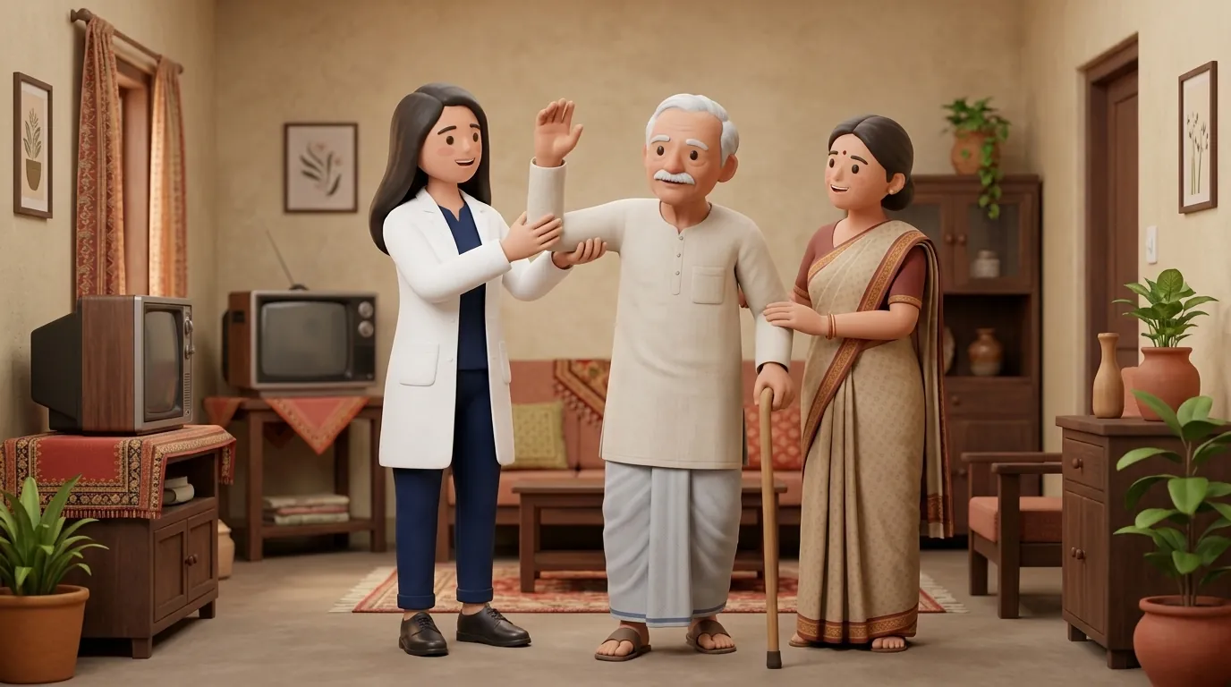
Achieving community ambulation in Indian residential environments requires managing practical architectural hurdles: ascending steep domestic stairs without handrails, stepping over raised bathroom thresholds, and boarding high public transport steps. An individual with L1-L3 paraplegia has the motor capacity to walk with bilateral elbow crutches or walking sticks. Domestic rehabilitation must focus heavily on stair climbing mechanics, teaching the patient to lead with the stronger leg during ascent and descending with crutches first, while ensuring living spaces are free of loose cotton carpets or clutter that could catch dropped toes.
Clinical Biomechanics and Neurological Recovery
The primary biomechanical requirement for upright walking is knee stability during stance phase. When quadriceps power is grade 3/5 or higher, the patient can extend the knee against gravity, but stance phase knee collapse remains a danger if the knee flexes beyond 15 degrees under full body weight. Ground Reaction Ankle-Foot Orthoses (GRAFOs) resolve this challenge through biomechanical ground reaction force manipulation. By holding the ankle in slight plantarflexion (approximately 2 to 3 degrees) and featuring a rigid anterior pre-tibial shell, the ground reaction force vector is directed anterior to the knee joint centre during stance, mechanically forcing the knee into extension without requiring high quadriceps effort.
Functional Movement Analysis and Clinical Assessment
Clinical examination rigorously measures manual muscle testing of key myotomes: hip flexors (L2, iliopsoas), knee extensors (L3, quadriceps), and ankle dorsiflexors (L4, tibialis anterior). The patellar tendon reflex is evaluated (often hyporeflexic or absent in lower motor neuron root lesions, or hyperreflexic in upper motor neuron conus lesions). Gait analysis evaluates stance stability: the clinician observes for knee hyperextension (genu recurvatum) resulting from weak quadriceps or excessive forward pelvic lurching resulting from absent gluteus maximus hip extensors.
Targeted Rehabilitation and Functional Independence Protocol
Gait rehabilitation progresses through structured stages. In Stage 1, closed-chain quadriceps strengthening is practiced on the mat table: partial terminal knee extensions and bridging drills. In Stage 2, sit-to-stand transitions are mastered from chairs of varying heights without using arm push-offs. In Stage 3, reciprocal overground walking is initiated: the patient swings the leg forward using active iliopsoas hip flexion, while the rigid AFO maintains foot clearance and prevents toe drag. Walking progresses from a four-point gait with a wheeled walker to a reciprocal gait using two forearm crutches, advancing to outdoor community walking.
Closed-Chain Step-Up and Eccentric Quadriceps Control Drill
Objective: Strengthen active L3 quadriceps motor units for stair climbing and stance stability
- 1Stand facing a sturdy 4-inch wooden exercise step or low wooden chowki with forearm crutches firmly placed.
- 2Lift your affected foot using active hip flexion, placing the entire sole flat on the step.
- 3Lean your body weight slightly forward over the step, loading the anterior thigh.
- 4Push downward through your foot, actively straightening your knee to lift your body onto the step.
- 5Hold the top position for 2 seconds while maintaining level pelvis alignment.
- 6Slowly and smoothly lower your trailing leg back to the floor over 3 controlled seconds.
| Orthotic Device | Biomechanical Mechanism | Clinical Indication Based on Muscle Power |
|---|---|---|
| Knee-Ankle-Foot Orthosis (KAFO) | Locks knee in full extension; rigid uprights span thigh to foot | Indicated when quadriceps strength is Grade 0 to 2/5 |
| Ground Reaction AFO (GRAFO) | Rigid anterior tibial shell directs ground reaction force anterior to knee | Indicated when quadriceps strength is Grade 3 to 4/5 with knee buckling |
| Solid Ankle AFO (Polypropylene) | Holds ankle at 90 degrees; prevents foot drop and mediolateral tilt | Indicated when quadriceps power is Grade 4/5 or 5/5 but foot drop persists |
| Posterior Leaf Spring AFO | Flexible calf shell assists toe clearance during swing phase | Indicated in high recovery where mild foot drop is the only residual deficit |
Red Flags and Emergency Escalation
During gait training with weak quadriceps, watch for persistent genu recurvatum (knee hyperextension) during stance phase. Allowing the patient to snap the knee backward to lock the joint stretches the posterior knee capsule, leading to severe chronic joint deformities and severe pain. Orthotic knee stops or GRAFO adjustments must be made immediately. In addition, inspect insensate skin over the anterior tibial crest and malleoli after every walking session: rigid plastic orthoses can cause deep pressure necrosis within hours if padding is inadequate.
Frequently Asked Questions
How do I know when to transition from heavy calipers (KAFOs) to lightweight AFOs?
The transition from full-leg KAFO calipers to below-knee AFOs becomes clinically viable when your voluntary knee extensor (quadriceps) strength achieves at least grade 3+ or 4 out of 5 against gravity and resistance. Your neuro-physiotherapist will conduct objective stance stability tests to ensure your knee will not buckle before modifying your orthotic prescription.
How do I book a home neuro-physiotherapist for orthotic gait retraining?
Under the NCAHP Act 2021, registered physiotherapists in India can be consulted directly at home without needing a doctor prescription. On BookPhysio.in, find licensed clinicians specialising in spinal cord injury gait retraining and pay directly at your home consultation via UPI or cash.
Related recovery guides & articles
BookPhysio.in Editorial Team
The BookPhysio.in editorial team writes and maintains this content, checking it against reliable clinical sources. Content follows our medical review policy: general information only, and no page claims a clinical review until a named registered physiotherapist has signed it off.
Read our medical review policy