
L4 to S1 Paraplegia Stepping Progression: Foot Drop Management and Plantarflexion
In short
Clinical management of foot drop and impaired plantarflexion in L4 to S1 paraplegia restores efficient, reciprocal community walking.
In this guide (6 sections)
Spinal lesions affecting the lower lumbar and sacral segments (L4 through S1) spare the primary motor power of the hip flexors, quadriceps, and hamstrings, preserving high functional ambulation potential. However, the selective neurological deficits striking the distal lower extremities present distinct functional impairments. Involvement of the L4 and L5 roots paralyzes the tibialis anterior, extensor hallucis longus, and peroneus longus, causing flaccid foot drop and severe loss of ankle eversion stability. Concomitant involvement of the S1 root denervates the gastrocnemius-soleus complex, abolishing terminal stance push-off. Managing this presentation requires specialised orthotic engineering paired with targeted gait retraining.
Domestic and Cultural Indian Realities
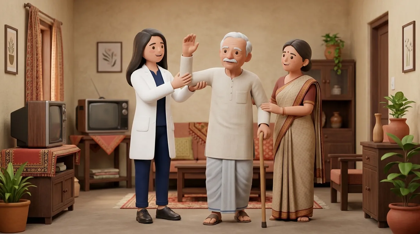
In Indian society, walking without shoes inside homes, temples, and social gatherings is customary. However, an individual with L4-S1 paraplegia walking barefoot with foot drop experiences severe instability: the toes catch on rugs or stone door sills, frequently leading to forward tripping falls. Furthermore, insensate soles across the S1-S2 dermatomes mean that stepping on small stones, dropped hairpins, or hot veranda tiles causes painless puncture lacerations and burns that can progress to osteomyelitis. Patients must adopt indoor orthopaedic footwear with integrated dynamic toe-lift orthoses, strictly avoiding traditional open-toe chappals or thin flip-flops that slide off paralyzed feet.
Clinical Biomechanics and Neurological Recovery
Normal human locomotion requires two distinct ankle actions: swing phase dorsiflexion to provide toe clearance, and terminal stance plantarflexion push-off to propel the body forward. In L4-S1 paraplegia, loss of dorsiflexion forces the patient into a compensatory steppage gait, exaggerating hip and knee flexion to lift the foot clear of the ground. In addition, loss of the calf muscle plantarflexion brake allows the tibia to collapse forward uncontrolled during mid-to-late stance, forcing the knee into excessive flexion and demanding high quadriceps energy. A dynamic carbon-composite energy-storing AFO solves both problems: holding the foot in neutral during swing and flexing during stance to deliver an elastic mechanical push-off.
Functional Movement Analysis and Clinical Assessment
Detailed manual muscle testing charts distal motor power: ankle dorsiflexion (L4, tibialis anterior), great toe extension (L5, extensor hallucis longus), and ankle plantarflexion (S1, gastrocnemius). The Achilles tendon reflex is graded (typically absent in S1 lesions). Gait kinematics are assessed over a 10-meter walkway: measuring cadence, step symmetry, and the presence of foot slap at initial contact. Proprioception and cutaneous sensation across the lateral foot and sole are mapped to identify insensate areas vulnerable to footwear ulceration.
Targeted Rehabilitation and Functional Independence Protocol
Rehabilitation focuses on reciprocal gait retraining and orthotic optimisation. Patients are fitted with custom carbon-fiber dynamic AFOs or lightweight articulated polypropylene AFOs with dorsiflexion-assist ankle joints. Overground gait drills emphasize three key movement phases: controlled heel strike (eliminating foot slap), mid-stance tibial progression, and explosive hip extension to maximize forward propulsion. Patients practice navigating uneven surfaces, climbing kerbs, and performing rapid direction changes without looking down at their feet.
Single-Limb Stance and Dynamic Step-Through Balance Drill
Objective: Train stance stability and trailing-limb hip extension in the presence of distal ankle weakness
- 1Stand tall beside a sturdy table or parallel bars with your hands hovering 1 inch above the surface.
- 2Shift your body weight fully onto your affected leg wearing the custom AFO.
- 3Maintain an upright posture, keeping your stance knee straight without snapping it backward.
- 4Swing your opposite leg smoothly forward past the stance foot in a natural walking step.
- 5Land softly on the forward heel, shift weight forward, and immediately step back to starting position.
- 6Focus on keeping your hips level and avoiding excessive lateral trunk sway.
| Orthotic Category | Biomechanical Impact | Clinical Suitability in L4-S1 Deficits |
|---|---|---|
| Carbon-Fiber Dynamic Energy-Return AFO | Stores kinetic energy during stance; releases explosive toe-off spring | Ideal for active community ambulators; highly durable and lightweight |
| Articulated AFO with Tamarack Flexure Joints | Allows free ankle dorsiflexion while providing elastic toe-lift assist | Excellent for negotiating stairs and inclines; requires good mediolateral stability |
| Solid-Ankle Polypropylene AFO | Completely immobilizes the ankle joint at 90 degrees | Used when severe ankle instability, spasticity, or genu recurvatum coexists |
| Soft Elastic Foot-Drop Cuff (Boxia) | Elastic tension strap attaches shoe laces to calf band | Comfortable indoor solution, but provides zero mediolateral or plantarflexion control |
Red Flags and Emergency Escalation
Due to S1 sensory loss covering the heel and plantar surface of the foot, patients are at extreme risk of developing painless neuropathic pressure ulcers from footwear seams or tight orthotic shells. Inspect both feet twice daily using a mirror: any blister, redness, or skin callosity mandates immediate orthotic modification. In addition, watch for recurring ankle inversion sprains: walking without adequate lateral orthotic support on uneven Indian streets can cause ligamentous tears or occult fibular fractures that go completely unfelt.
Frequently Asked Questions
Will I need to wear an ankle-foot orthosis (AFO) permanently for foot drop?
This depends on the degree of nerve root regeneration. In incomplete lower motor neuron cauda equina injuries, peripheral nerves regenerate slowly over 12 to 24 months. If voluntary tibialis anterior power recovers to grade 4/5 or higher, the orthosis can often be weaned off. However, if motor recovery plateaus with residual foot drop, continuing to wear a lightweight carbon AFO prevents tripping falls and preserves joint alignment.
How do I arrange in-home gait assessment and orthotic fitting with a physiotherapist?
Under the NCAHP Act 2021, registered physiotherapists in India can be engaged directly for home gait evaluations. Through BookPhysio.in, you can book an experienced physiotherapist who will analyze your walking kinematics, recommend optimal AFO designs, and train balance in your domestic setting. Fees are paid directly to the clinician at your home appointment.
Related recovery guides & articles
BookPhysio.in Editorial Team
The BookPhysio.in editorial team writes and maintains this content, checking it against reliable clinical sources. Content follows our medical review policy: general information only, and no page claims a clinical review until a named registered physiotherapist has signed it off.
Read our medical review policy