
Orthostatic Hypotension in Acute SCI: Verticalisation and Abdominal Binders
In short
Orthostatic hypotension significantly delays early rehabilitation in spinal cord injury; structured verticalization protocols restore hemodynamic stability.
In this guide (6 sections)
Orthostatic hypotension (OH) is an exceptionally common and disabling cardiovascular complication following cervical and high thoracic spinal cord injury (injuries at or above T6). Defined clinically as a sustained reduction in systolic blood pressure of at least 20 mmHg or diastolic blood pressure of at least 10 mmHg within 3 minutes of assuming an upright posture, it causes profound cerebral hypoperfusion. Patients experience lightheadedness, dizziness, blurred vision, yawning, diaphoresis, and syncope (fainting) whenever attempting to sit upright in bed or transfer into a wheelchair. Understanding the underlying neurogenic vasomotor failure and implementing progressive verticalization protocols is essential to enable active physical rehabilitation.
Domestic and Cultural Indian Realities
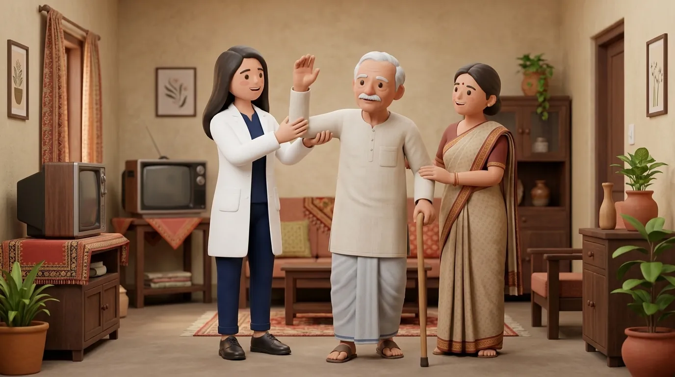
In Indian home environments, early orthostatic episodes often induce intense anxiety among family members. After witnessing a patient turn pale, lose consciousness, and slump over upon sitting, families frequently enforce prolonged, uninterrupted bed rest, keeping the patient flat for weeks or months. This prolonged immobilization creates a vicious cycle: bed rest induces cardiovascular deconditioning and reduces blood plasma volume, dramatically worsening orthostatic intolerance upon subsequent sitting attempts. Educating family caregivers to view dizziness as a manageable autonomic adjustment and equipping them with elastic abdominal binders and graduated compression stockings allows safe home mobilization.
Clinical Biomechanics and Neurological Recovery
In neurologically intact individuals, transitioning from supine to upright triggers baroreceptors in the carotid sinus and aortic arch, stimulating sympathetic outflow that causes reflex vasoconstriction in the splanchnic and lower limb vascular beds. In spinal cord injuries above T6, descending sympathetic vasomotor pathways originating in the medulla are disconnected from the sympathetic preganglionic neurons in the spinal cord (T1-L2). Gravity pulls blood downward, causing massive venous pooling in the compliant splanchnic mesenteric vascular bed and paralyzed lower extremities. Venous return drops precipitously, reducing stroke volume and cerebral blood pressure.
Functional Movement Analysis and Clinical Assessment
Clinical evaluation begins with serial orthostatic vital signs: measuring blood pressure and heart rate in supine after 5 minutes of rest, followed by measurements at 1, 3, and 5 minutes after ascent to 45 degrees, 60 degrees, and 90 degrees. Baseline resting blood pressure in chronic tetraplegia is typically low (around 90/60 mmHg); a drop below 70/40 mmHg induces immediate cerebral ischemia. The clinician monitors for classic presyncopal warning signs: pallor, yawning, diaphoresis around the forehead, and cognitive slowing.
Targeted Rehabilitation and Functional Independence Protocol
The non-pharmacological management protocol combines external mechanical compression with progressive postural acclimatisation. Step 1: Apply a wide, multi-panel elastic abdominal binder spanning from the lower rib cage to the iliac crests before raising the head; compressing the massive splanchnic reservoir accounts for up to 60 percent of orthostatic blood pressure support. Step 2: Don thigh-high graduated compression stockings (20 to 30 mmHg) to reduce lower limb pooling. Step 3: Utilize a progressive tilt table or reclining wheelchair, raising the backrest by 15-degree increments every 5 minutes while monitoring blood pressure, allowing the neuro-vascular system to re-establish autoregulatory tone.
Bedside Progressive Verticalisation and Upper Limb Pumping Routine
Objective: Promote central venous return and cerebral perfusion during postural transitions
- 1Fit your wide elastic abdominal binder firmly across your stomach while lying completely flat.
- 2raise the head of your hospital bed or stack firm pillows to raise your upper torso to 30 degrees.
- 3Perform slow, forceful bilateral shoulder shrugs and overhead arm reaching to stimulate venous return.
- 4Maintain the 30-degree ascent for 5 minutes, confirming complete absence of lightheadedness.
- 5Advance the bed ascent to 45 degrees, continuing active upper limb movement.
- 6Progressively advance toward 75 to 90 degrees upright sitting over a 20-minute structured session.
| Intervention Modality | Physiological Mechanism | Clinical Practicality and Effectiveness |
|---|---|---|
| Elastic Abdominal Binder | Compresses splanchnic mesenteric venous pool, raising preload | Highly effective; simple to apply; primary non-pharmacological defense |
| Thigh-High Compression Stockings | Exerts external pressure on calf and femoral venous plexuses | Effective, but challenging to don; requires daily skin checks |
| Progressive Tilt Table Verticalisation | Re-conditions cerebral autoregulatory vascular resistance | Gold standard for inpatient rehabilitation; highly structured |
| Oral Salt and Fluid Expansion | Increases circulating plasma volume and systemic hydration | Requires medical clearance to prevent fluid overload or kidney strain |
Red Flags and Emergency Escalation
If a patient loses consciousness (syncope) during verticalization, immediately recline the wheelchair fully or lower the bed flat while raising both lower extremities above heart level; cerebral perfusion will restore within seconds. Never leave an acutely lightheaded patient unattended in a standard upright wheelchair. In addition, distinguish orthostatic hypotension from autonomic dysreflexia: while hypotension causes low blood pressure and pallor upon standing, dysreflexia causes dangerous hypertension, severe pounding headache, and facial flushing. Furthermore, always remove abdominal binders when lying supine to prevent skin irritation and digestive disturbance.
Frequently Asked Questions
How tightly should an abdominal binder be worn for orthostatic hypotension?
The binder should be snug enough to exert firm, consistent compressive pressure across the entire abdomen from the lower ribs to the pubic bone, compressing the splanchnic blood vessels. However, it must not be so tight that it restricts deep diaphragmatic breathing or causes skin redness over the pelvic bones.
How do I schedule home physiotherapy for tilt table training and verticalization?
Under the NCAHP Act 2021, registered physiotherapists in India provide direct domiciliary neuro-rehabilitation without doctor prescriptions. On BookPhysio.in, book a qualified physiotherapist who will structure safe, monitored verticalization protocols in your home. Fees are paid directly to the clinician at your home appointment.
Related recovery guides & articles
BookPhysio.in Editorial Team
The BookPhysio.in editorial team writes and maintains this content, checking it against reliable clinical sources. Content follows our medical review policy: general information only, and no page claims a clinical review until a named registered physiotherapist has signed it off.
Read our medical review policy