
Partial Toe Amputation and Ray Resection: Balance Retraining and Pressure Transfer
In short
Amputation of a single toe or metatarsal ray alters the mechanics of the entire foot, transferring massive pressure to neighbouring joints. Learn how to prevent secondary transfer ulcers.
In this guide (6 sections)
Partial toe amputations and single-ray resections are among the most common surgical procedures performed to halt localized osteomyelitis or digital gangrene in diabetic patients. While removing an infected digit successfully controls sepsis, it permanently alters the transverse and longitudinal weight-bearing mechanics of the foot. When the hallux (great toe) or first metatarsal ray is removed, the primary anchor of the medial column is lost. Ground reaction forces instantly transfer laterally onto the second and third metatarsal heads, producing severe transfer lesions and secondary ulceration within months. Conversely, amputating lesser toes permits adjacent digits to drift, sublux, and develop painful deformities. Post-operative rehabilitation requires custom silicone digital prostheses, total contact orthotic insoles, and balance retraining to restore stability.
Domestic and Cultural Indian Realities
In Indian society, many patients view the loss of a single toe as a minor event that should not interfere with daily barefoot habits. However, walking barefoot across unyielding marble or tile floors after a toe amputation accelerates secondary breakdown. When the great toe is missing, the patient loses push-off power and balance, causing a pronounced limp and clumsy foot placement. To steady themselves, patients walk with an externally rotated foot, generating high shear stresses beneath the remaining lesser metatarsal heads. Furthermore, wearing traditional open chappals allows neighbouring toes to drift into the empty amputation space, creating severe friction and corns. Patients must wear custom-moulded medical footwear equipped with a silicone toe spacer that fills the surgical defect.
Clinical Biomechanics and Neuropathic Breakdown
The hallux normally bears up to twice the load of any lesser digit during the terminal stance and pre-swing phases of gait. When the hallux or first ray is resected, the foot loses the windlass mechanism and medial propulsive lever. Ground reaction forces shift abruptly to the second metatarsal head, where peak vertical pressures regularly surge past 800 kilopascals. Because the second metatarsal shaft is slender and rigidly fixed at the tarsometatarsal joint, this transferred overload causes rapid sub-metatarsal callus formation, stress fractures, and full-thickness ulceration. When a second toe is amputated, the hallux drifts into severe hallux valgus, while the third toe claws into the vacant gap.
Clinical Assessment and Neuropathy Screening
Assessment involves clinical inspection of the surgical site, verifying scar mobility, tissue healing, and the alignment of remaining digits. Dynamic baropodometric pressure mapping or Harris mat analysis identifies areas of transferred peak pressure under adjacent metatarsal heads. Range of motion of the ankle and midtarsal joints is evaluated, checking for compensatory equinus tightness. Sensory testing with a 10g monofilament maps the extent of neuropathy, and peripheral pulses are palpated to verify arterial perfusion.
Targeted Offloading and Intrinsic Foot Conditioning
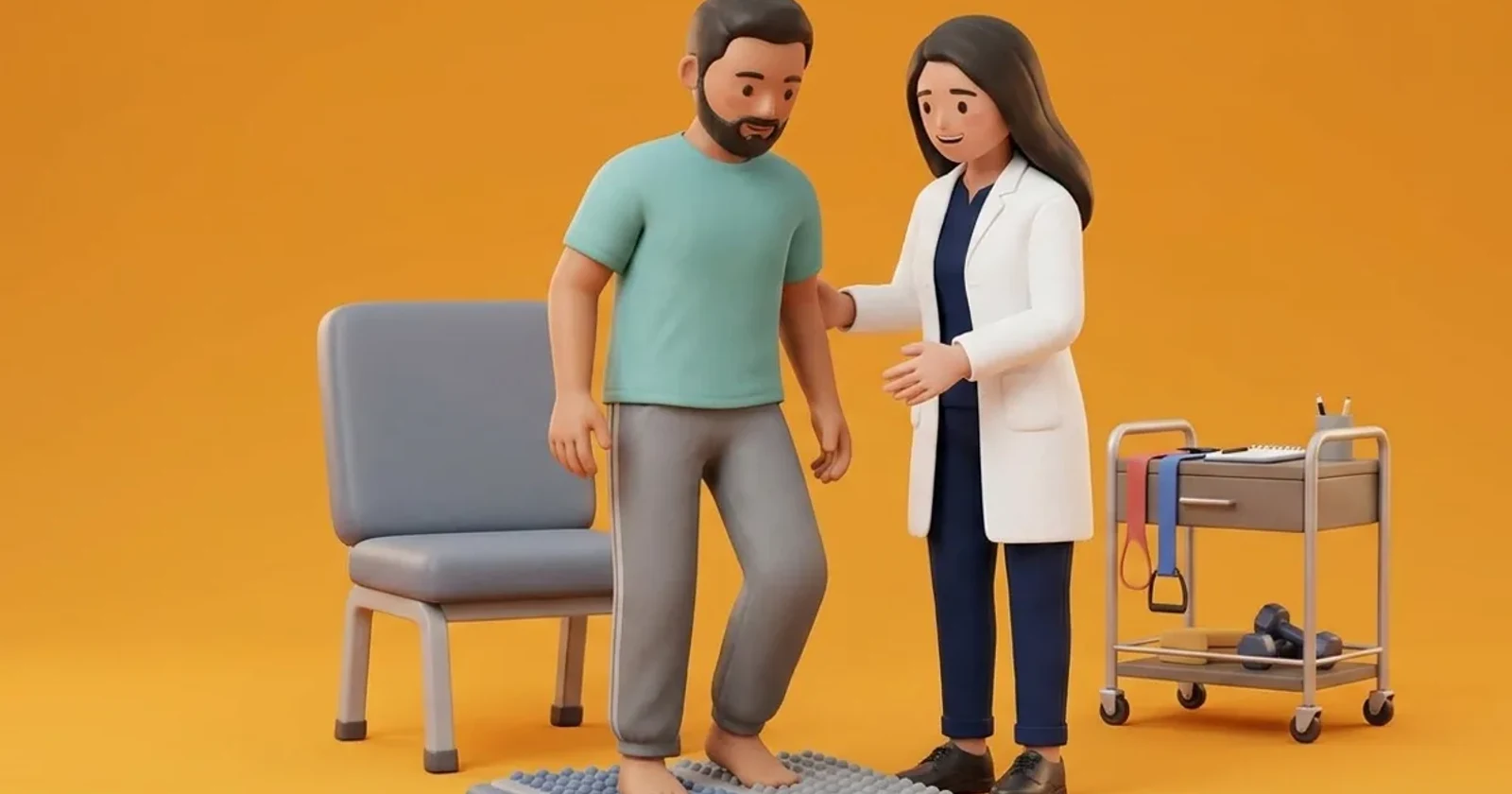
Rehabilitation focuses on custom orthotic fabrication to manage transferred mechanical loads. A custom silicone toe filler is moulded to occupy the empty space left by the amputated toe, preventing neighbouring digits from drifting and maintaining shoe alignment. The insole incorporates a metatarsal pad and relief wells beneath the second and third metatarsal heads. A rigid rocker-bottom sole is prescribed to roll the foot forward smoothly without relying on forefoot push-off. Physiotherapy incorporates balance drills and ankle stability exercises.
Seated Bilateral Ankle Eversion and Inversion Retraining
Objective: Strengthen ankle stabilisers to compensate for altered forefoot weight-bearing lines
- 1Sit securely in a firm chair with feet placed flat on a non-slip floor mat.
- 2Keeping your heels anchored firmly in place, gently lift the inner edges of both feet, rolling outward onto your outer foot borders.
- 3Hold the inverted position steadily for 3 seconds.
- 4Slowly roll feet flat, then gently lift the outer edges, pressing the inner borders down into eversion.
- 5Hold the everted position for 3 seconds, then return to neutral resting position.
| Amputation Site | Secondary Biomechanical Pressure Shift | Prescribed Clinical Orthotic Solution |
|---|---|---|
| Hallux (Great Toe) Amputation | Massive load transfer to 2nd and 3rd metatarsal heads; loss of push-off | Custom total contact insole with 2nd MTP relief well; rigid rocker sole shoe |
| First Metatarsal Ray Resection | Complete medial column collapse; severe overload of lesser metatarsals | Rigid carbon shank footplate; deep medial arch support; rocker-bottom sole |
| Second Toe Amputation | Severe hallux valgus drift; 3rd toe migration into empty gap | Medical-grade silicone toe spacer filler; broad square toe box footwear |
| Fifth Metatarsal Ray Resection | Loss of lateral column support; compensatory medial roll-off and shear | Lateral column wedge build-up; high-top heel counter ankle support |
Red Flags and Urgent Diabetic Foot Escalation
Immediate medical escalation is necessary if a patient discovers redness, calluses, or blood blisters beneath the remaining metatarsal heads or along the surgical scar. Spreading erythema, localized heat, cloudy exudate, or a sudden increase in foot swelling indicates secondary infection requiring emergency podiatric review. Rapid intervention prevents adjacent toe amputations.
Frequently Asked Questions
Why do I need a silicone toe filler if only one toe was removed?
When a toe is missing, the empty space inside your shoe allows the remaining toes to slide, drift sideways, and curl. This drift creates severe friction corns and causes your shoe to fold in awkwardly. A silicone toe filler maintains the natural shape of your shoe and keeps your remaining toes straight.
Why am I developing a thick callus under my second toe after losing my big toe?
Your big toe normally bears the greatest amount of weight when you walk. Without it, your body automatically dumps that heavy pressure onto the neighbouring second toe bone, which is not designed for such loads. The thick callus is an urgent warning sign of extreme pressure that will turn into an ulcer without custom insole offloading.
Related recovery guides & articles
BookPhysio.in Editorial Team
The BookPhysio.in editorial team writes and maintains this content, checking it against reliable clinical sources. Content follows our medical review policy: general information only, and no page claims a clinical review until a named registered physiotherapist has signed it off.
Read our medical review policy