
Pelvic Floor sEMG Biofeedback: Coordination vs Strength Retraining in India
In short
Many people perform Kegels incorrectly without realizing it. Discover how real-time surface electromyography (sEMG) biofeedback retrains neuromuscular coordination and relaxation.
In this guide (6 sections)
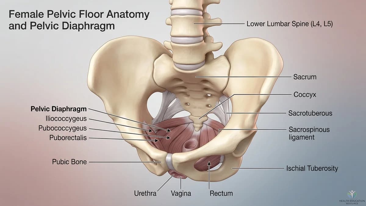
Pelvic floor muscle training is widely recommended for urinary incontinence, organ prolapse, and pelvic pain, yet clinical studies reveal that over 50 percent of individuals perform pelvic contractions incorrectly when guided by verbal or written instructions alone. In India, patients frequently hold their breath, clench their gluteals, or paradoxically bear down as if having a bowel movement, actions that worsen incontinence and strain pelvic ligaments. Surface electromyography (sEMG) biofeedback bridges this gap by converting invisible neuromuscular electrical activity into real-time visual and auditory signals on a clinical monitor. This objective feedback allows patients to retrain motor coordination, verify muscle relaxation, and build functional strength.
1. Functional Biomechanics: Motor Unit Action Potentials and sEMG Waveforms
Surface EMG biofeedback records the summation of motor unit action potentials (MUAPs) generated during pelvic floor muscle activation using specialized internal vaginal/rectal sensors or perianal surface electrodes. The clinical monitor displays key physiological parameters: resting baseline tone (normal is under 2.0 microvolts), peak voluntary contraction amplitude, contraction rise time (speed of motor unit recruitment), contraction stability (endurance holding), and post-contraction return to baseline. In hypertonic pelvic floor disorders, baseline resting tone remains elevated (3 to 8 microvolts), demonstrating that the muscle cannot relax. In hypotonic weakness, peak amplitude is blunted. Biofeedback teaches patients both how to contract and, crucially, how to fully relax.
2. Domestic Indian Context: Daily Chores, Ergonomics, and Privacy
In Indian clinical settings, many patients feel apprehensive about pelvic instrumentation due to modesty and unfamiliarity with medical technology. Explaining that sEMG biofeedback is entirely passive, recording electrical signals produced by the patient's own muscles rather than administering an electrical shock, provides immediate reassurance. Learning how to isolate the pelvic floor without substituting accessory muscles (like the adductors or rectus abdominis) transforms daily home exercise into an effective, targeted routine that can be practiced quietly in any household setting.
3. Clinical Special Tests and Physical Examination
A standard sEMG assessment protocol follows the Glazer Protocol: (1) 1-minute pre-baseline rest, (2) 5 rapid 'flick' contractions, (3) 5 sustained 10-second endurance contractions, (4) a 60-second endurance hold, and (5) a 1-minute post-baseline rest to assess muscle fatigue and recovery rate.
| Protocol Stage | Electrophysiological Target | Normal Clinical Value | Dysfunctional Finding |
|---|---|---|---|
| 1. Resting Baseline | Resting muscle tone at complete rest | Below 2.0 microvolts (uV) | Elevated baseline (>3 uV) indicating spasm |
| 2. Fast-Twitch Flicks | Explosive recruitment & rapid relaxation | Rapid peak spike with return to baseline | Sluggish onset or delayed muscle release |
| 3. Endurance Holds | 10-second sustained contractions | Stable plateau with <15% muscle decay | Rapid muscle fade, trembling, early fatigue |
| 4. Post-Test Baseline | Complete recovery after fatigue protocol | Returns cleanly to <2.0 uV | Failure to relax; elevated muscle contracture |
4. Stage-by-Stage Functional Physiotherapy Protocol
Rehabilitation begins by calibrating visual biofeedback, eliminating accessory muscle substitution, and transferring coordination into functional upright tasks.
Phase 1: Visual Biofeedback Calibration and Resting Drop Protocol
Objective: Achieve consistent resting baseline tone below 2.0 microvolts before attempting active strengthening.
- 1With the sensor comfortably in place, view the real-time line graph on the clinical monitor.
- 2Take slow diaphragmatic breaths, observing how the signal line responds to breathing.
- 3Focus your attention on dropping the line into the green zone (below 2.0 microvolts).
- 4Notice the physical sensation of pelvic release and softening that corresponds with the low signal on the screen.
- 5Maintain this quiet baseline for 30 seconds before attempting any active contraction.
Phase 2: Target-Matching Fast-Twitch and Endurance Contractions
Objective: Match voluntary muscle recruitment to visual target boxes on the biofeedback monitor.
- 1Watch the screen as a target box appears at a designated amplitude (for example, 15 microvolts).
- 2On an exhalation, contract your pelvic floor smoothly to guide the signal line directly into the target box.
- 3Hold the line steadily inside the target for 8 seconds without letting it fluctuate or fall.
- 4Release immediately when the target ends, watching the line drop cleanly back below baseline within 1 second.
- 5This builds precise neurological motor control rather than uncoordinated clenching.
Phase 3: Dual-Channel Accessory Muscle De-Coupling
Objective: Eliminate unwanted abdominal or gluteal clenching during pelvic floor activation.
- 1Surface electrodes are placed over your abdominal wall while the pelvic sensor records internally.
- 2Attempt a pelvic floor contraction while keeping the abdominal signal line completely flat.
- 3If your abdominal line spikes, reduce your effort until only the pelvic line rises.
- 4This de-couples the pelvic floor from superficial muscle substitution, building true isolated control.
Discontinue biofeedback and seek medical consultation if you experience: sudden severe pelvic pain during sensor placement, fresh vaginal or rectal bleeding, signs of local mucosal infection or fever, or acute inability to pass urine.
5. Clinic Consultation, Home Visits, and Transparent Fee Benchmarks
Under the National Commission for Allied and Healthcare Professions (NCAHP) Act 2021, patients in India can consult a registered pelvic health physiotherapist directly without an upfront referral, though BookPhysio.in does not ask for one. Specialized clinics in major Indian cities feature computerized sEMG biofeedback workstations in private, clinical rooms. Clinic consultations typically range between ₹500 and ₹1,500, with home visits ranging from ₹700 to ₹2,000 where portable units are available. Patients pay their physiotherapist directly at each consultation, with zero platform commission.
6. Frequently Asked Questions
Does sEMG biofeedback hurt or give an electric shock?
No, sEMG biofeedback is completely painless and non-invasive. The sensor acts purely as a listening device that detects the natural microvolt electrical signals generated by your own muscle fibres when you contract. It does not deliver any electrical current or shock.
How many biofeedback sessions are typically needed for pelvic retraining in India?
Most patients require 4 to 8 supervised clinical biofeedback sessions spaced over 6 to 10 weeks to master correct motor isolation and down-training. Once correct muscle recruitment and relaxation are established, patients continue an independent home maintenance exercise programme.
Related recovery guides & articles
BookPhysio.in Editorial Team
The BookPhysio.in editorial team writes and maintains this content, checking it against reliable clinical sources. Content follows our medical review policy: general information only, and no page claims a clinical review until a named registered physiotherapist has signed it off.
Read our medical review policy