
Posterior Canal BPPV: Canalithiasis vs Cupulolithiasis and Dix-Hallpike Diagnosis
In short
Posterior semicircular canal BPPV accounts for the vast majority of mechanical vertigo cases; distinguishing free-floating canalithiasis from cupular cupulolithiasis determines whether an Epley or Semont manoeuvre succeeds.
In this guide (8 sections)
Posterior Semicircular Canal Mechanics and Otoconia Dynamics
Benign Paroxysmal Positional Vertigo (BPPV) of the posterior semicircular canal represents approximately 85 to 90 percent of all otolith displacement presentations encountered in neuro-otology practice. Calcium carbonate crystals (otoconia) detach from the gelatinous matrix of the utricular macula and migrate into the gravity-dependent posterior canal. The clinical presentation diverges fundamentally based on whether these displaced particles remain free-floating within the canal lumen (canalithiasis) or become physically adhered to the cupular cupula (cupulolithiasis).
In canalithiasis, head motion in the plane of the canal produces hydrodynamic fluid drag (endolymph flow), deflecting the cupula after a distinct 1 to 5 second latency period. This induces paroxysmal rotational vertigo accompanied by an upbeat, torsional nystagmus beating toward the dependent ear that resolves within 30 to 60 seconds as the particles settle. Conversely, in cupulolithiasis, the cupula becomes directly gravity-sensitive; positional testing produces immediate, non-fatiguing upbeat torsional nystagmus that persists for as long as the head remains in the provocative position.
Domestic Context: Vulnerabilities in Indian Daily Life
In Indian households, posterior canal BPPV is frequently triggered during ordinary domestic routines that involve rapid changes in vertical head orientation. Common provocations include bending forward to fill water buckets from low bathroom taps, bowing down during morning pooja rituals or prostrating on the floor (sashtang pranam), looking up at overhead attic lofts (chhajjas) or ceiling fans, and turning over rapidly on firm cotton mattresses.
A sudden spinning attack in a wet tiled Indian bathroom poses severe risks of slip-and-fall injuries against concrete or marble thresholds. Family members often mistake acute positional vertigo for an impending stroke or severe cervical spondylosis, prompting unnecessary brain scans or unhelpful neck collars. Accurate bedside canal identification prevents prolonged disability and allows rapid particle clearance without invasive interventions.
Clinical Testing: The Dix-Hallpike Diagnostic Protocol
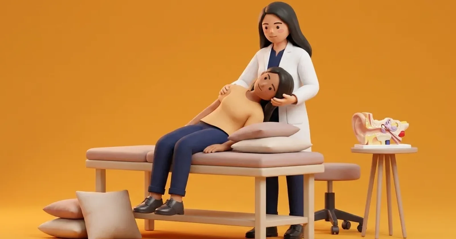
The gold-standard diagnostic procedure is the Dix-Hallpike test. The patient is seated longways on an examination couch, the head is rotated 45 degrees toward the test ear, and the clinician rapidly moves the patient into a supine position with the neck extended 20 to 30 degrees below the horizontal plane. The clinician observes the eyes for characteristic nystagmus features under Frenzel lenses or infrared video goggles.
- Latency Period: A delay of 1 to 5 seconds before nystagmus onset confirms canalithiasis, whereas immediate onset without latency indicates cupulolithiasis.
- Nystagmus Vector: Mixed upbeat and geotropic torsional nystagmus, where the top poles of the eyes beat toward the dependent, downwards-facing ear.
- Duration and Fatigue: Crescendo-decrescendo nystagmus lasting under 60 seconds indicates canalithiasis; non-fatiguing nystagmus lasting beyond 60 seconds points to cupular adhesion.
- Reversal on Sitting: Returning the patient upright reverses endolymph flow, producing a brief, mild downbeat and reverse torsional nystagmus.
Stage-by-Stage Functional Repositioning Protocol
- Stage 1: Mechanical Canal Clearance: Perform a verified Epley canalith repositioning manoeuvre for canalithiasis, pausing 30 to 60 seconds at each 90-degree rotational station. For cupulolithiasis, execute a brisk Semont liberating manoeuvre to mechanically detach cupular otoconia using rapid inertial deceleration.
- Stage 2: Post-Manoeuvre Activity Guidelines: Avoid extreme prolonged head extension or aggressive inversion for the initial 24 hours. Modern evidence refutes rigid cervical collar use; normal gentle functional head movements are encouraged once acute canal clearing is achieved.
- Stage 3: Vestibular Habituation and Balance Re-education: If mild residual floating sensations persist despite negative repeat Dix-Hallpike testing, introduce Brandt-Daroff habituation exercises and dynamic standing balance drills on foam surfaces.
Clinical Exercise Protocol: Home Particle Settling Exercise
Modified Brandt-Daroff Habituation Protocol
Objective: Disperses residual microscopic otoconial debris and promotes central vestibular adaptation following successful canal repositioning.
- 1Sit upright on the edge of a firm bed or plinth with your feet resting flat on the floor.
- 2Turn your head 45 degrees to the left, then lie down briskly on your right side while keeping your head turned; maintain this position for 30 seconds or until any spinning subsides.
- 3Return to the upright seated position smoothly and remain still for 30 seconds to allow endolymph stabilisation.
- 4Turn your head 45 degrees to the right, lie down quickly on your left side, and hold for 30 seconds before returning to the upright seated position.
Clinical Comparison: Canalithiasis vs Cupulolithiasis
| Clinical Parameter | Canalithiasis Variant | Cupulolithiasis Variant |
|---|---|---|
| Otoconia Location | Free-floating within the posterior canal lumen | Adhered directly to the gelatinous cupula |
| Dix-Hallpike Latency | Present (1 to 5 seconds delay) | Absent (immediate onset of nystagmus) |
| Duration of Nystagmus | Transient (less than 60 seconds) | Persistent (greater than 60 seconds) |
| Primary Treatment Manoeuvre | Epley canalith repositioning manoeuvre | Semont liberating manoeuvre (inertial deceleration) |
| Fatigability on Repeat Testing | Marked fatigue on repeated positioning | Minimal or absent fatigability |
Red Flags and HINTS Stroke Differentiation
A comprehensive examination must rule out central neurological pathology. Positional vertigo with downbeating nystagmus without a torsional component, direction-changing nystagmus without position changes, or persistent spontaneous nystagmus while seated upright are red flags for cerebellar or brainstem lesions. Under the HINTS exam (Head Impulse, Nystagmus, Test of Skew), an acute central stroke is suspected if the head impulse test is normal in a patient with spontaneous vertigo, if gaze-evoked nystagmus changes direction, or if vertical skew deviation is visible on alternate cover testing. Patients presenting with ataxia, dysarthria, or facial weakness require immediate emergency medical escalation.
Frequently Asked Questions
Why does dizziness sometimes persist after a successful Epley manoeuvre?
Residual dizziness occurs in about one-third of patients due to transient utricular dysfunction, microscopic otoconial dust remaining in the canal, or central sensory recalibration delays. It typically responds well to gentle walking and Brandt-Daroff habituation.
Can I perform an Epley manoeuvre on myself at home without clinical assessment?
Self-treatment without diagnostic confirmation carries risks of misdiagnosis or canal switching, where particles inadvertently enter the horizontal or anterior canals. A registered physiotherapist should first verify the exact canal and variant.
Related recovery guides & articles
BookPhysio.in Editorial Team
The BookPhysio.in editorial team writes and maintains this content, checking it against reliable clinical sources. Content follows our medical review policy: general information only, and no page claims a clinical review until a named registered physiotherapist has signed it off.
Read our medical review policy