
Sensorimotor Retraining for Diabetic Neuropathy: Textured Surfaces and Visual Cues
In short
Sensory re-education stimulates remaining tactile pathways and cortical representation in diabetic neuropathy. Discover structured protocols using textured surfaces and visual compensation.
In this guide (6 sections)
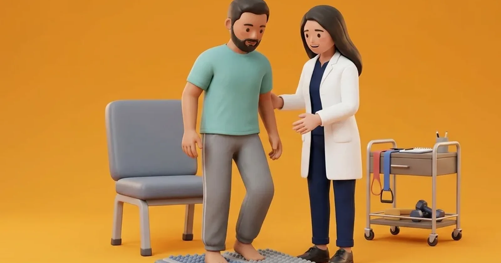
Sensory re-education is a neurorehabilitation paradigm designed to stimulate remaining peripheral mechanoreceptors and preserve cortical somatosensory representation in patients with diabetic polyneuropathy. As peripheral axonal degeneration progresses, central sensory maps in the primary somatosensory cortex undergo disorganization due to diminished afferent input. Structured sensory training using graded tactile surfaces, textured sensory mats, and conscious visual-tactile pairing helps maximize the sensitivity of surviving nerve fibres. When integrated with visual compensation techniques, sensorimotor training improves dynamic balance, improves foot placement awareness during walking, and reduces the frequency of stumbling episodes.
Domestic and Cultural Indian Realities
In Indian households, the transition from bare floors to sensory retraining requires strict safety supervision. While healthy individuals benefit from walking on acupressure mats or pebble stones, applying rough, unyielding textures to an insensate diabetic foot can cause severe skin lacerations and ulcers. Traditional acupressure wooden boards or spike mats sold in local markets must never be used by individuals with diabetic neuropathy. Instead, sensory re-education must utilize safe, medical-grade elastomeric mats with smooth, rounded silicone nodules or soft fabrics (such as velvet, terrycloth, and fine corduroy) used in a seated position. This ensures sensory stimulation without the risk of skin puncture or high mechanical shear.
Clinical Biomechanics and Neuropathic Breakdown
Cutaneous mechanoreceptors in the plantar sole (Meissner corpuscles, Merkel discs, Ruffini endings, and Pacinian corpuscles) provide high-resolution feedback regarding ground texture, slip velocity, and shear direction. In diabetes, axonal atrophy and demyelination raise tactile thresholds, meaning only large, coarse stimuli can evoke a cortical response. By presenting distinct, controlled textures while the patient actively looks at the foot, neuroplastic cross-modal integration occurs: visual observation primes the somatosensory cortex, reinforcing weak peripheral signals. This conscious feedback loop helps the central nervous system interpret muted plantar inputs more effectively during functional walking.
Clinical Assessment and Neuropathy Screening
Assessment begins by establishing baseline sensory perception using 10g monofilaments and quantitative two-point discrimination testing on the plantar hallux and heel. The clinician evaluates joint position sense (proprioception) at the first metatarsophalangeal joint by moving the toe up or down in small 5-degree increments with the patient eyes closed. Dynamic gait analysis observes foot drop, high-steppage gait, or heavy slapping footfalls, which indicate that the patient is relying on auditory cues to confirm ground contact.
Targeted Offloading and Intrinsic Foot Conditioning
Rehabilitation exercises are always performed in a seated, non-weight bearing posture to prevent skin pressure damage. The patient moves the sole of the foot across various safe textures placed on a floor board, identifying smooth, ridged, and soft surfaces while watching the motion. Training advances to visual line tracking, where the patient practices walking along high-contrast floor lines while wearing protective, flexible-soled shoes, using deliberate heel-to-toe foot placement guided by eye focus.
Seated Multi-Texture Tactile Recognition Drill
Objective: Stimulate plantar cutaneous mechanoreceptors safely without weight-bearing pressure
- 1Sit securely in a comfortable chair under bright room lighting with bare feet.
- 2Place two different soft fabric swatches (such as a soft microfibre towel and a smooth silk scarf) flat on the floor side by side.
- 3Gently slide the sole of your right foot back and forth across the microfibre towel for 30 seconds while looking directly at your foot.
- 4Next, move your foot onto the smooth silk surface and slide it gently, noting the difference in texture visually and mentally.
- 5Repeat the sensory gliding sequence with your left foot.
- 6Inspect the bottom of both feet thoroughly in your mirror after the session to ensure zero redness.
| Material / Surface Type | Tactile Property & Safety Rating | Clinical Usage Recommendation |
|---|---|---|
| Soft Rounded Silicone Nodular Mat | Medical-grade elastomeric nodules; completely safe | Use in seated position only; provides tactile stimulation without shear |
| Graded Fabric Swatches (Cotton / Fleece) | Smooth, low-friction textures; completely safe | Ideal for visual-tactile discrimination drills while seated |
| Commercial Wooden Acupressure Spikes | Sharp, rigid, high point pressure; EXTREMELY HAZARDOUS | Strictly prohibited; causes puncture wounds and deep plantar ulcers |
| Natural River Pebble Walkways | Uneven, unyielding, high shear stress; DANGEROUS | Avoid barefoot walking; causes contusions and Charcot joint trauma |
Red Flags and Urgent Diabetic Foot Escalation
Immediate medical escalation is necessary if the patient observes any redness, abrasion, blister, or skin peeling following sensory practice. Any sudden decrease in joint position awareness, new muscle twitching, or sudden unilateral foot weakness requires clinical reassessment to exclude acute radiculopathy or peroneal nerve compression.
Frequently Asked Questions
Can acupressure foot mats improve nerve sensation in diabetes?
No. Acupressure mats with hard plastic or wooden points are extremely dangerous for diabetic feet. Because neuropathy prevents you from feeling pain, these sharp points can pierce your skin, cause deep bruising, and trigger severe non-healing ulcers. Always use soft, flat, medical-grade textures in a seated position.
How does looking at my feet help my balance when walking?
When nerve damage blunts the feeling in your feet, your brain cannot tell where your feet are in space. By looking consciously at your step placement and floor obstacles, your visual cortex substitutes for your damaged proprioceptive nerves, allowing you to place your feet smoothly and safely.
Related recovery guides & articles
BookPhysio.in Editorial Team
The BookPhysio.in editorial team writes and maintains this content, checking it against reliable clinical sources. Content follows our medical review policy: general information only, and no page claims a clinical review until a named registered physiotherapist has signed it off.
Read our medical review policy