
Pseudogout (CPPD) Knee Synovitis: Gentle Joint Rehabilitation Protocols
In short
Calcium pyrophosphate dihydrate (CPPD) crystal shedding into the knee joint triggers acute inflammatory synovitis mimicking septic arthritis. Discover evidence-based joint decompression, quadriceps reactivation, and safe mobility protocols.
In this guide (6 sections)
Calcium pyrophosphate dihydrate (CPPD) crystal deposition disease, historically termed pseudogout, is a common microcrystalline arthropathy predominantly affecting older adults. CPPD crystals deposit within articular cartilage and fibrocartilage (such as the knee menisci), a radiographic phenomenon known as chondrocalcinosis. When crystals shed from cartilage into the synovial fluid cavity, they trigger an explosive acute inflammatory synovitis that most commonly targets the knee joint. The joint becomes rapidly distended with a massive synovial effusion, presenting with severe throbbing pain, warmth, and complete loss of weight-bearing tolerance. Following acute medical aspiration, structured physiotherapy is vital to evacuate residual oedema, counteract arthrogenic muscle inhibition, and restore joint mechanics.
Domestic and Cultural Indian Realities
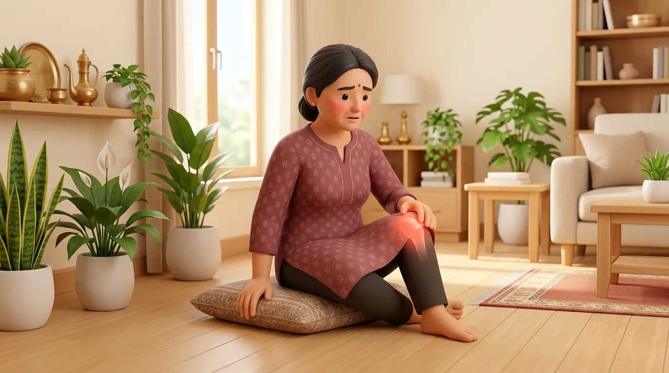
In Indian homes, an acute episode of pseudogout in an elderly family member often develops abruptly following minor systemic stress, such as a mild viral infection, a long car journey, or dehydration during hot weather. Because the knee swells rapidly and becomes hot, family members frequently misinterpret it as a simple sprain and apply vigorous hot mustard oil massage, which dramatically worsens synovial hyperemia and intra-articular pressure. Patients cannot bend the knee to sit on low stools, use Indian squat commodes, or climb domestic stairs. Families must provide an over-toilet commode chair, install supportive handrails, raise the leg on pillows, and avoid all heat applications until the acute crystal inflammatory flare subsides.
Clinical Biomechanics and Inflammatory Pathology
The pathogenesis of CPPD involves abnormal pyrophosphate metabolism, where excessive extracellular inorganic pyrophosphate binds with calcium within the cartilage matrix. Crystal shedding into the joint space induces neutrophil recruitment through Toll-like receptor and inflammasome activation. The resulting massive joint effusion increases intra-capsular hydrostatic pressure. Mechanoreceptors within the distended capsule trigger reflex arthrogenic muscle inhibition (AMI), shutting down motor unit recruitment in the vastus medialis and rectus femoris. Attempting heavy resisted exercises during this acute phase accelerates cartilage wear and increases patellofemoral friction.
Clinical Assessment and Functional Staging
Clinical evaluation begins by grading knee joint effusion using the stroke or bulge test for minor fluid or patellar tap testing for massive effusions. Joint line tenderness, local skin calor, and active range of motion are systematically recorded. Radiographs of the knee reveal linear, punctate calcification along the articular cartilage and within the menisci (chondrocalcinosis). Synovial fluid analysis under polarized light microscopy confirms rhomboid-shaped, weakly positively birefringent crystals, ruling out septic arthritis and urate gout.
Targeted Mobility and Joint Preservation Exercises
Rehabilitation focuses on non-weight bearing rhythmic motion to facilitate synovial fluid resorption, combined with sub-maximal isometric quadriceps setting.
Supine Active-Assisted Heel Slides with Towel Cradle
Objective: Evacuate intra-articular fluid and restore knee flexion-extension range without loading the joint
- 1Lie flat on your back on a firm bed with your legs straight and head supported by a pillow.
- 2Loop a long towel or bedsheet under the sole of your affected foot, holding the ends in both hands.
- 3Gently pull on the towel to slide your heel smoothly toward your buttocks, bending your knee to a comfortable, pain-free limit.
- 4Pause for 3 seconds, then slowly slide your heel back down straight, exhaling smoothly.
| Clinical Factor | Acute Pseudogout Flare Phase | Post-Inflammatory Recovery Phase |
|---|---|---|
| Effusion Management | Joint aspiration by rheumatologist; cold pack application and elevation | Active muscle pump contractions and retrograde lymphatic massage |
| Quadriceps Training | Sub-maximal isometric quadriceps setting at 20 degrees flexion with zero resistance | Progressive closed-chain mini-squats, step-ups, and seated knee extension |
| Ambulation Support | Axillary or elbow crutches maintaining partial or non-weight bearing status | Independent unassisted walking with supportive, cushioned footwear |
| Domestic Transfers | Raised toilet seat (19 inches) and raised dining chair with armrests | Gradual return to normal household activities, avoiding deep floor squatting |
Red Flags and Immediate Rheumatology Escalation
An acutely hot, swollen, painful knee joint accompanied by systemic chills, rigors, high fever, or skin pustules requires emergency medical hospitalization to rule out septic arthritis. A joint infected with bacteria can present identically to pseudogout and can destroy articular cartilage within 24 to 48 hours if left undrained.
Frequently Asked Questions
Can knee exercises dissolve CPPD crystals or reverse chondrocalcinosis?
No. Exercise cannot dissolve calcium pyrophosphate crystals or reverse chondrocalcinosis within the cartilage. However, evidence-based physiotherapy helps manage morning stiffness, evacuates joint effusion, reverses quadriceps inhibition, and restores stable walking mechanics.
How do I schedule a home visit by a qualified physiotherapist for knee rehabilitation via BookPhysio.in?
Under the National Commission for Allied and Healthcare Professions (NCAHP) Act 2021, Indian patients enjoy direct access to registered physiotherapists without an obligatory doctor referral. On BookPhysio.in, you schedule an appointment with a verified clinician in your city. Payment is made directly to the therapist at your home appointment via cash or UPI; BookPhysio.in does not process booking fees online.
Related recovery guides & articles
BookPhysio.in Editorial Team
The BookPhysio.in editorial team writes and maintains this content, checking it against reliable clinical sources. Content follows our medical review policy: general information only, and no page claims a clinical review until a named registered physiotherapist has signed it off.
Read our medical review policy