
Real-Time Ultrasound (RTUS): Transabdominal Pelvic Floor Visualization and Biofeedback in India
In short
Visualizing your pelvic floor muscles moving on an ultrasound screen ensures you are lifting rather than bearing down. Learn how transabdominal RTUS guides recovery.
In this guide (6 sections)
Transabdominal Real-Time Ultrasound (RTUS) has revolutionized pelvic health physiotherapy by providing a non-invasive, comfortable, and objective method to assess pelvic floor muscle function. For many patients in India, including young women, postpartum mothers, men recovering from prostate surgery, and elderly individuals, internal digital vaginal or rectal examinations can feel intimidating or culturally sensitive. Transabdominal RTUS places an ultrasound probe gently over the lower abdomen, allowing patients to watch their bladder base elevate in real time as their pelvic floor contracts. This visual confirmation eliminates guesswork so patients learn to lift their pelvic floor rather than accidentally bearing down.
1. Functional Biomechanics: Cranial Bladder Base Displacement and Muscle Synergy
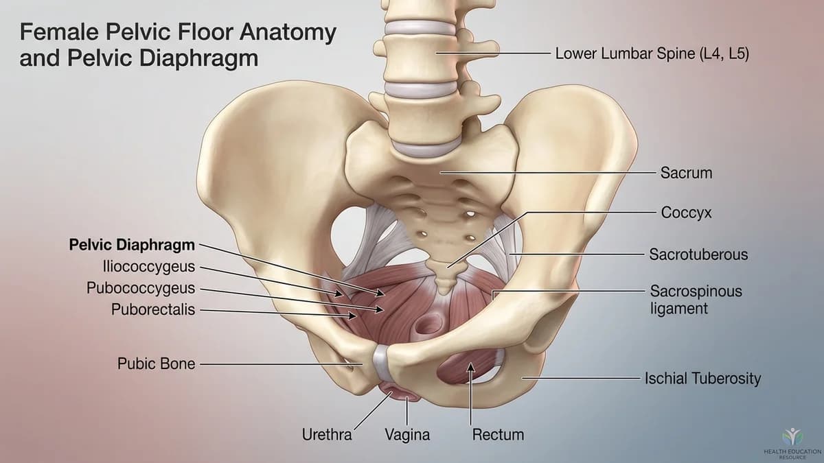
The pelvic floor and abdominal wall function as an integrated muscular unit. When the probe is placed transversely in the midline just superior to the symphysis pubis, the acoustic beam visualizes the hypoechoic, fluid-filled bladder lumen, with the pelvic floor resting directly beneath the bladder base. During a correct voluntary pelvic floor contraction, the levator ani elevates cranially (inward and upward), producing a distinct upward displacement of the bladder floor on the monitor screen (typically 5 to 10 millimetres). If a patient performs an incorrect Valsalva bearing down action, the bladder base drops caudally (downward), providing instant visual proof of maladaptive technique.
2. Domestic Indian Context: Daily Chores, Ergonomics, and Privacy
In Indian society, cultural modesty often makes individuals reluctant to seek help for urinary incontinence or pelvic weakness because they dread internal physical examinations. Transabdominal RTUS offers an ideal, respectful solution: the patient remains fully clothed, exposing only the lower abdomen just above the waistband. Furthermore, seeing their own muscles moving on a screen provides immediate confidence to patients who doubted whether they possessed the ability to control their pelvic floor, making it an empowering educational tool in Indian clinics.
3. Clinical Special Tests and Physical Examination
Assessment requires a comfortably full bladder (consuming 500 to 750 millilitres of water 1 hour prior to testing). A 3.5 to 5.0 MHz curved array probe is angled 15 to 30 degrees caudally. The clinician measures bladder base displacement in millimetres, assesses contraction velocity, and tests muscle endurance during a sustained 10-second hold and during a simulated cough.
| Assessment Metric | Normal Correct Contraction | Dysfunctional Bearing Down | Clinical Action Required |
|---|---|---|---|
| Bladder Base Vector | Cranial lift upward by 5 to 10+ mm | Caudal descent downward (>2 mm) | Immediate cueing to stop bearing down |
| Deep Core Synergy | Transversus abdominis firms without doming | Rectus abdominis bulges, breath held | Re-train diaphragmatic breathing coordination |
| Cough Reflex Response | Instant anticipatory upward lift | Bladder base pushed sharply downward | Train 'The Knack' pre-contraction drill |
| Patient Biofeedback | Visual confirmation reinforces correct motor map | Visual correction breaks harmful habit | Use screen for progressive target holds |
4. Stage-by-Stage Functional Physiotherapy Protocol
The protocol progresses from visual biofeedback calibration to motor consolidation, weaning off the screen as proprioceptive accuracy improves.
Phase 1: Real-Time Visual Biofeedback Calibration on RTUS
Objective: Establish an accurate mental map of pelvic floor elevation using live ultrasound visualization.
- 1Lie comfortably on the treatment plinth with knees bent and your gaze directed at the ultrasound monitor.
- 2Locate your bladder on the screen, a dark triangular or rounded shape with a bright floor line beneath it.
- 3Inhale gently into your belly. As you exhale, imagine gently pulling your tailbone toward your pubic bone.
- 4Watch the screen: observe the bright line at the base of the bladder lift upward toward your head.
- 5If the line moves downward, stop immediately, take a relaxed breath, and try a lighter, gentler upward lift.
Phase 2: Sustained Ultrasound Target Holds and Endurance Training
Objective: Build slow-twitch postural muscle endurance while confirming zero muscle fade on screen.
- 1Initiate a clean cranial lift of the bladder base on the ultrasound monitor.
- 2Maintain the elevation at the peak height for 8 seconds while breathing continuously out loud.
- 3Observe the screen: verify that the bladder base does not slowly sag back downward during the hold.
- 4Release the contraction smoothly, watching the bladder base return cleanly to its resting level.
- 5Rest for 10 seconds before the next repetition.
Phase 3: Screen Blinded Proprioceptive Verification
Objective: Verify that correct motor recruitment continues when visual feedback is removed.
- 1Turn your head away from the ultrasound screen so you cannot see the image.
- 2Perform what you feel is a correct upward pelvic floor lift and hold for 5 seconds.
- 3The physiotherapist monitors the screen to verify that a true cranial lift occurred without abdominal substitution.
- 4Look back at the screen to confirm your success, transitioning confidence from visual feedback to internal body feel.
If real-time ultrasound reveals unexpected incidental findings, such as a large solid pelvic mass, severe bladder wall thickening with irregular mucosal lesions, or bilateral hydronephrosis (kidney swelling), the physiotherapist will immediately refer you to a urologist or gynaecologist for urgent medical evaluation.
5. Clinic Consultation, Home Visits, and Transparent Fee Benchmarks
Under the National Commission for Allied and Healthcare Professions (NCAHP) Act 2021, patients in India can consult a registered pelvic health physiotherapist directly without an upfront referral, though BookPhysio.in does not ask for one. RTUS assessments are performed in modern outpatient clinics equipped with dedicated diagnostic ultrasound apparatus. Clinic sessions across Indian cities range between ₹500 and ₹1,500, with home visits ranging from ₹700 to ₹2,000 where available. Patients pay their physiotherapist directly at each consultation, with zero platform commission.
6. Frequently Asked Questions
Is real-time ultrasound safe during pregnancy in India?
Yes, transabdominal ultrasound uses completely safe, non-ionizing high-frequency sound waves with zero radiation risk to either the mother or developing baby. It is widely used throughout pregnancy to evaluate pelvic floor function and assess diastasis recti separation safely.
Why do I need a full bladder for transabdominal pelvic ultrasound?
Sound waves travel easily through fluid. A comfortably full bladder acts as an acoustic window, pushing intestinal gas out of the way and providing clear contrast so the physiotherapist can visualize the movement of the pelvic floor muscles resting beneath the bladder base.
Related recovery guides & articles
BookPhysio.in Editorial Team
The BookPhysio.in editorial team writes and maintains this content, checking it against reliable clinical sources. Content follows our medical review policy: general information only, and no page claims a clinical review until a named registered physiotherapist has signed it off.
Read our medical review policy