
Rheumatoid Wrist Extensor Tendon Rupture Prevention: Caput Ulnae Syndrome and Splinting
In short
Chronic rheumatoid synovitis at the distal radioulnar joint triggers caput ulnae syndrome, risking devastating extensor tendon attrition ruptures. Master preventive splinting, forearm biomechanics, and tendon protection rules.
In this guide (6 sections)
In rheumatoid arthritis, the wrist joint is involved in over 90 percent of patients, serving as a primary driver of upper extremity disability. Persistent synovitis within the distal radioulnar joint (DRUJ) destroys the triangular fibrocartilage complex (TFCC) and attenuates dorsal radioulnar ligaments, allowing the distal ulnar head to subluxate dorsally. This clinical entity, known as caput ulnae syndrome, presents with a prominent, ballotable ulnar head (piano key sign) and painful forearm rotation. Crucially, the scalloped, eroded dorsal ulnar bone acts as an abrasive, sharp saw against the overlying extensor tendons within the fourth and fifth extensor compartments. Repeated friction leads to progressive extensor tendon attrition rupture, initiating at the small finger and marching radially toward the index finger (Vaughn-Jackson syndrome).
Domestic and Cultural Indian Realities
Indian domestic tasks place heavy repetitive rotational demands on the wrist and distal radioulnar joint. Wringing wet heavy cotton sarees and bedsheets by hand, churning lentils with a traditional wooden hand churner (mathani), and turning stiff brass water taps generate extreme supination-pronation torques. In the presence of a dorsally subluxated ulnar head, these forceful twisting motions saw the extensor digitorum communis and extensor digiti minimi tendons directly across jagged bony spurs. Homemakers must immediately cease manual wringing, use tap turners or lever taps, adopt electric kitchen appliances, and wear rigid thermoplastic resting splints during sleep and functional wrist supports during domestic activities.
Clinical Biomechanics and Inflammatory Pathology
The distal radioulnar joint relies on soft tissue restraints for stability, particularly the TFCC and the dorsal and palmar radioulnar ligaments. In rheumatoid disease, proliferative synovial pannus infiltrates the extensor retinaculum and DRUJ capsule, causing ligamentous elongation. The carpus subluxates volarly and shifts ulnarly, while the ulnar head springs dorsally. Tendon rupture occurs through a dual mechanism: inflammatory tenosynovitis induces intrinsic collagen necrosis and localized tendon ischemia, while mechanical friction against the sharp, eroded ulnar head causes mechanical attrition. Once the extensor digiti minimi ruptures, the extensor digitorum slips to the ring and middle fingers follow in rapid succession if surgical or orthotic intervention is delayed.
Clinical Assessment and Functional Staging
Physiotherapists examine the wrist for the piano key sign: pressing firmly downward on the prominent ulnar head elicits pain and demonstrates vertical depression followed by immediate elastic rebound. Forearm supination and pronation range of motion are quantified using a goniometer. Active digital extension is carefully tested: patients are asked to extend each finger individually at the metacarpophalangeal joint while the wrist is held in neutral. Tendon integrity is confirmed by evaluating tenodesis action: passively flexing the wrist should naturally produce passive extension of all fingers.
Targeted Mobility and Joint Preservation Exercises
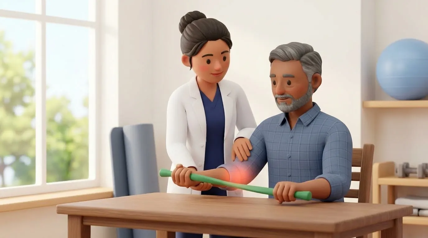
Rehabilitation requires strict avoidance of forced forearm pronation-supination under load. Exercise focuses on active-assisted digital tendon gliding with the wrist immobilized in 15 to 20 degrees of extension.
Neutral Wrist-Supported Digital Extensor Tendon Gliding
Objective: Maintain digital extensor tendon excursion and prevent adhesions without inducing abrasive friction over the ulnar head
- 1Rest your affected forearm flat on a table, supported on a rolled towel with your wrist resting in slight extension (15 degrees) and palm facing downward.
- 2Slowly lift all four fingers off the table together, straightening them at the knuckle joints while keeping your wrist still.
- 3Hold the extended position gently for 3 seconds without forcing backward hyperextension.
- 4Slowly lower the fingers back to touch the table surface smoothly without snapping.
| Clinical Intervention | Protective Joint Strategy | Dangerous High-Attrition Pattern |
|---|---|---|
| Splinting Strategy | Custom-molded dorsal blocking wrist splint maintaining 15 degrees extension and neutral forearm | Unsplinted heavy use or flexible elastic bandages that compress the ulnar head into tendons |
| Domestic Chores | Spin-dry washing machines; utilizing pump bottles and wide lever handles | Manual wringing of wet laundry and twisting tight jar lids with naked hands |
| Manual Exercises | Gentle isolated tendon glides with supported neutral wrist alignment | Aggressive wrist circumduction, heavy dumbbell curls, and forceful grip squeezes |
| Surgical Referral Timing | Immediate surgical consult upon piano key pain to consider Darrach or Sauve-Kapandji procedure | Waiting until multiple fingers drop before seeking orthopaedic hand surgery review |
Red Flags and Immediate Rheumatology Escalation
Sudden, painless loss of active extension in the small finger (drop finger), followed by inability to extend the ring finger, represents an acute extensor tendon rupture. This is a surgical emergency; the patient must be referred immediately to an orthopaedic hand surgeon for tendon transfer or grafting before adjacent tendons rupture.
Frequently Asked Questions
Can physiotherapy reattach or heal a ruptured extensor tendon?
No. Once a tendon has completely ruptured from mechanical attrition, physiotherapy exercises cannot reconnect the torn tendon ends; surgical reconstruction or tendon transfer by an orthopaedic hand surgeon is mandatory. Physiotherapy plays a critical role in prevention through early splinting and postoperative rehabilitation to restore hand function.
How do I book an experienced hand therapist for home visits via BookPhysio.in?
Under the National Commission for Allied and Healthcare Professions (NCAHP) Act 2021, Indian patients enjoy direct access to registered physiotherapists without an obligatory physician referral. On BookPhysio.in, you schedule an appointment with a verified clinician in your city. You pay the consultation fee directly to the therapist at your home appointment via cash or UPI; BookPhysio.in collects no booking fees online.
Related recovery guides & articles
BookPhysio.in Editorial Team
The BookPhysio.in editorial team writes and maintains this content, checking it against reliable clinical sources. Content follows our medical review policy: general information only, and no page claims a clinical review until a named registered physiotherapist has signed it off.
Read our medical review policy