
Sphenopalatine Ganglion Neuralgia: Craniofacial Tension and Cervical Release
In short
Sphenopalatine ganglion neuralgia causes deep retro-orbital facial pain, nasal congestion, and upper cervical tension. Targeted physiotherapy targets suboccipital restrictions and trigeminocervical convergence to soothe pain.
In this guide (7 sections)
Sphenopalatine ganglion (pterygopalatine ganglion) neuralgia produces intense, deep-seated burning or aching facial pain centred behind the eye, root of the nose, and upper jaw. The condition is frequently accompanied by prominent autonomic signs, including ipsilateral eye watering (lacrimation), conjunctival redness, and nasal congestion. Because sensory inputs from the upper cervical spine and trigeminal nerve converge within the trigeminocervical nucleus in the brainstem, chronic suboccipital tension and C1-C2 segmental dysfunction can amplify pain signaling. Physiotherapy addresses this craniofacial circuit through upper cervical mobilization, suboccipital release, and gentle autonomic calming techniques.
Domestic and Cultural Indian Realities
Environmental triggers in Indian homes frequently aggravate sphenopalatine-related pain. Sleeping directly beneath a high-speed ceiling fan or facing direct air conditioning drafts in offices provokes cold-induced vasoconstriction and neural irritation across the face. Prolonged forward head posture while studying for competitive exams or working long hours on laptops adds sustained mechanical tension to the suboccipital musculature. In Indian domestic life, taking a warm, quiet shower, avoiding direct drafts to the head, and practicing gentle nasal breathing help soothe the sensitive cranial nerves.
Clinical Biomechanics and Anatomical Healing
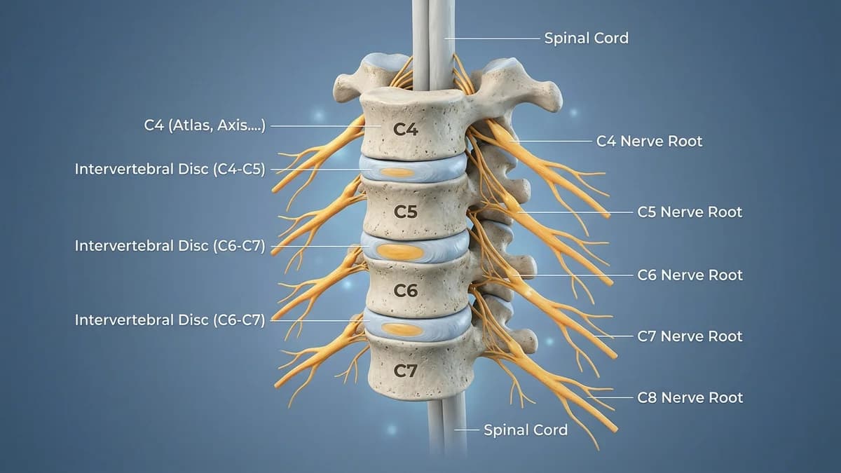
The sphenopalatine ganglion sits in the pterygopalatine fossa, communicating with the maxillary branch of the trigeminal nerve and autonomic fibers of the vidian nerve. Nociceptive inputs from the upper cervical spine (specifically C1, C2, and C3 spinal nerves) project to the caudal portion of the spinal trigeminal nucleus. Consequently, mechanical restriction or muscle spasm in the suboccipital triangle (rectus capitis posterior and obliquus capitis) sensitizes trigeminal pathways, lowering the threshold for craniofacial neural flares. Relieving suboccipital tightness reduces this central afferent barrage.
Clinical Assessment and Diagnostic Examination
Clinical evaluation combines a detailed cranial nerve screening with an upper cervical biomechanical examination. The physiotherapist performs the Flexion-Rotation Test (FRT) to assess rotational mobility at the C1-C2 motion segment. Digital palpation across the suboccipital muscles, greater occipital nerve groove, and superficial temporalis checks for trigger points that reproduce facial ache. Autonomic signs such as eye tearing and nasal blockage are documented to differentiate the condition from primary trigeminal neuralgia or cluster headaches.
Targeted Rehabilitation and Mobility Exercises
Rehabilitation emphasizes manual suboccipital release, gentle axial cervical traction, and diaphragm-focused autonomic down-regulation. Patients learn self-release techniques using two soft rubber balls placed under the base of the skull, combined with slow nasal diaphragmatic breathing to support parasympathetic tone.
Suboccipital Decompression with Soft Ball Release
Objective: Relieves upper cervical tension to dampen trigeminocervical afferent sensitivity.
- 1Lie on your back on a comfortable mat with your knees bent and feet flat on the floor.
- 2Place two soft tennis balls or massage balls tied in a sock directly under the base of your skull (just above the neck).
- 3Allow the weight of your head to rest gently on the balls without pressing forcefully.
- 4Inhale slowly through your nose for 4 seconds, feeling your belly rise, and exhale gently for 6 seconds.
- 5Maintain this relaxing pressure for 3 to 5 minutes, keeping your jaw relaxed and teeth separated.
Rehabilitation Milestones and Recovery Timeline
| Stage and Duration | Primary Objective | Clinical Intervention | Target Milestone | |
|---|---|---|---|---|
| Stage 1 (Weeks 0-2) | Autonomic soothing and trigger reduction | Draft protection, breathing drills, suboccipital rest | Reduction in daily facial burning intensity | |
| Stage 2 (Weeks 2-5) | Upper cervical mobilization and muscle release | Therapist C1-C2 mobilization and soft tissue release | Symmetrical cervical rotation to at least 40 degrees on FRT | |
| Stage 3 (Weeks 5-8) | Deep neck flexor and postural retraining | Cervical stabilization and scapular alignment | Marked decrease in autonomic eye watering and headache frequency | |
| Stage 4 (Weeks 8-12) | Long-term resilience and self-management | Home ball release, ergonomic habits, stress control | Sustained symptom stability and restored daily comfort |
Red Flags and Specialist Escalation
Seek emergency medical and neurological care if severe facial or eye pain is accompanied by pupil asymmetry (Horner syndrome), sudden drooping of the eyelid, double vision, thunderclap headache, or acute loss of facial sensation.
Frequently Asked Questions
Can treating the back of my neck really help relieve pain behind my eye?
Yes, nerves from the upper neck and sensory nerves from the face converge at the trigeminocervical nucleus in the brainstem. Releasing tension and restoring mobility in the upper neck reduces the neurological irritability that projects pain forward into the eye and temple.
How does breathing through the nose help with craniofacial neuralgia?
Slow, rhythmic nasal breathing stimulates parasympathetic nervous pathways and engages the diaphragm. This down-regulates systemic sympathetic overactivity, helping ease involuntary facial muscle clenching and vascular sensitivity.
Related recovery guides & articles
BookPhysio.in Editorial Team
The BookPhysio.in editorial team writes and maintains this content, checking it against reliable clinical sources. Content follows our medical review policy: general information only, and no page claims a clinical review until a named registered physiotherapist has signed it off.
Read our medical review policy