
Transmetatarsal Amputation Rehabilitation: Achilles Lengthening and Shoe Fillers
In short
Transmetatarsal amputation preserves the heel and ankle but disrupts muscular balance and the forefoot lever arm. Discover clinical rehabilitation protocols and prosthetic shoe fillers.
In this guide (6 sections)
Transmetatarsal amputation (TMA) is a valuable limb salvage procedure performed to eradicate forefoot gangrene, extensive deep phlegmon, or severe osteomyelitis while preserving the calcaneus, talus, and midfoot joints. Although a TMA retains the patient natural heel pad and allows limited barefoot household transfers, it dramatically alters the structural biomechanics of the foot. Transecting the metatarsal shafts detaches the insertions of the tibialis anterior, extensor digitorum longus, and peroneus brevis, while leaving the powerful gastrocnemius-soleus complex and tibialis posterior intact. This severe muscular imbalance pulls the residual foot into equinovarus contracture, tilting the stump downward and inward. Structured post-amputation rehabilitation, combined with percutaneous Achilles tendon lengthening (TAL), custom shoe fillers, and rigid rocker soles, is vital to prevent stump ulceration.
Domestic and Cultural Indian Realities
Following a TMA, Indian patients frequently assume that preserving the heel means they can return to walking barefoot at home. However, walking barefoot on a shortened residual foot causes intense peak shear and compressive forces directly across the sharp distal metatarsal bone ends and surgical scar. When stepping barefoot onto hard marble or stone floors, the shortened foot behaves like a peg leg, concentrating immense pressure on the plantar-distal stump margin and causing rapid surgical wound breakdown. In Indian homes, patients must wear custom indoor shoes equipped with full-length carbon footplates and soft silicone toe fillers at all times. Rising from floor sitting or low charpoys must be modified to prevent scraping the distal stump against the floor.
Clinical Biomechanics and Neuropathic Breakdown
The loss of the forefoot metatarsal parabola abolishes the third rocker (forefoot rocker) of the gait cycle, reducing the mechanical lever arm required for forward propulsion by approximately 50 percent. Without an intact forefoot lever, the Achilles tendon acts with reduced mechanical advantage, pulling the residual foot into equinus. The remaining plantarflexor muscles overpower the weakened dorsiflexors, causing the distal metatarsal stump to point toward the floor during stance. Peak vertical pressures over the distal metatarsal stumps exceed 900 kilopascals. To restore normal gait, the prosthetic prescription must feature a full-length rigid carbon shank that re-establishes the missing forefoot lever arm, paired with a custom elastomer toe filler that prevents the foot from sliding forward inside the shoe.
Clinical Assessment and Neuropathy Screening
Assessment begins with surgical wound inspection, evaluating scar healing, skin graft adherence, and tissue mobility over the bone ends. Ankle joint passive range of motion is measured with a goniometer to identify early equinus or varus contracture. Sensation is mapped with 10g monofilaments across the residual stump and plantar heel. Vascular status is verified through palpation of the posterior tibial pulse and Doppler ABI. This confirms adequate blood supply to support tissue healing under mechanical loading.
Targeted Offloading and Intrinsic Foot Conditioning
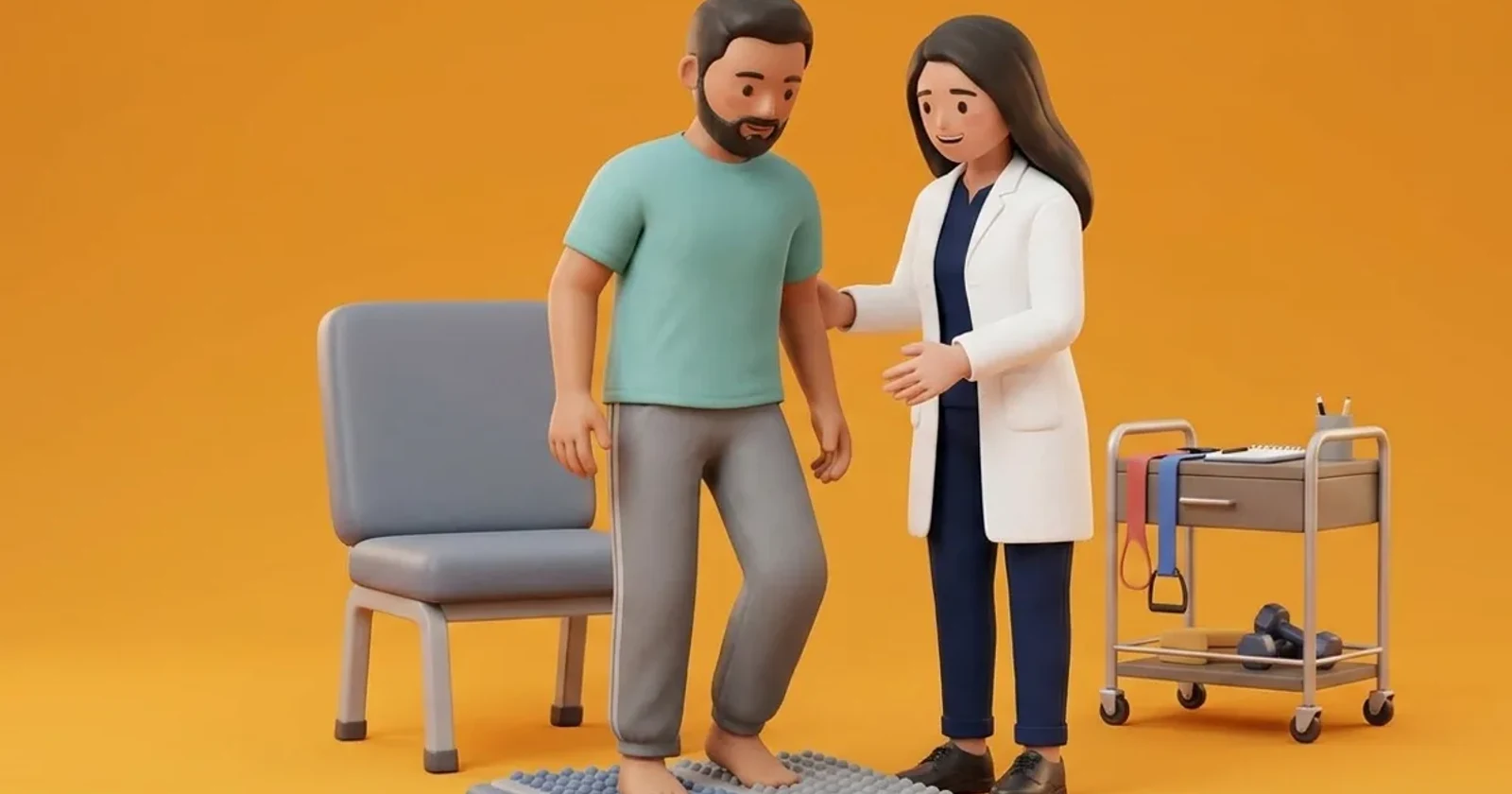
Rehabilitation progresses from non-weight bearing stump conditioning to progressive weight-bearing in a rigid rocker shoe. Physiotherapy emphasizes gentle passive ankle dorsiflexion stretching and active tibialis anterior strengthening to counter equinus forces. Once the surgical incision has matured, a custom total contact orthosis with an integrated lightweight silicone toe filler is fabricated. Gait retraining teaches a shortened step length, smooth weight transfer across the rocker sole, and avoidance of sudden push-off maneuvers.
Seated Passive Residual Foot Dorsiflexion Stretch
Objective: Maintain ankle dorsiflexion and prevent equinovarus contracture of the TMA stump
- 1Sit comfortably on your bed with the amputated limb extended straight in front of you.
- 2Loop a wide, non-elastic cotton towel or strap around the distal end of your residual foot.
- 3Keeping your knee straight, gently pull the strap backward toward your chest.
- 4Draw the stump into neutral dorsiflexion until you feel a gentle stretch in the back of your calf.
- 5Hold the sustained stretch for 20 to 30 seconds without bouncing.
- 6Slowly release tension and rest for 10 seconds before repeating.
| Prosthetic Component | Engineering Specification | Clinical Biomechanical Function |
|---|---|---|
| Custom Moulded Insole with Toe Filler | Multi-density EVA base with soft silicone or Plastazote toe block | Fills empty anterior shoe space; prevents residual stump from sliding forward |
| Full-Length Rigid Carbon Footplate | Lightweight stiff carbon fibre plate extending full shoe length | Replaces missing forefoot lever arm; restores forward roll-over stability |
| External Rigid Rocker-Bottom Sole | Curved sole with apex placed just proximal to metatarsal stump ends | Eliminates propulsive push-off stress; smooths forward transition |
| High-Top Ankle Support Collar | Padded leather collar extending above the malleoli with firm lacing | Controls subtalar varus tilting; prevents heel slippage inside shoe |
Red Flags and Urgent Diabetic Foot Escalation
Immediate medical escalation is necessary if a patient discovers redness, blisters, or skin breakdown along the distal surgical scar or beneath the bony stump ends. A prominent bone edge poking against thin skin indicates imminent ulceration. Any purulent discharge, localized warmth, offensive odor, or systemic fever demands emergency surgical review to prevent ascending infection into the tarsal joints.
Frequently Asked Questions
Why do many patients have an Achilles tendon lengthening procedure during TMA surgery?
Transecting the metatarsal bones removes the muscles that pull the foot upward, while leaving the strong calf muscles intact. Without an Achilles lengthening, the calf muscle pulls the residual foot downward into a severe pointed position (equinus), forcing the fresh bone ends into the ground. Lengthening the Achilles tendon balances the foot and protects the surgical stump.
Can I walk barefoot inside the house for quick trips after a transmetatarsal amputation?
No. Walking barefoot on a TMA stump is extremely dangerous. Without toes to spread your weight, every step places tremendous pressure directly onto the cut bone ends and delicate surgical scar. Stepping barefoot on hard floors will quickly cause ulcers and bone infection. Always wear your prescription shoe with its custom toe filler indoors.
Related recovery guides & articles
BookPhysio.in Editorial Team
The BookPhysio.in editorial team writes and maintains this content, checking it against reliable clinical sources. Content follows our medical review policy: general information only, and no page claims a clinical review until a named registered physiotherapist has signed it off.
Read our medical review policy