
Acetabular Fracture Post-ORIF: Hip Touch-Down Weight Bearing Progression
In short
Guide to postoperative acetabular fracture rehabilitation. Master touch-down weight bearing, hip motion precautions, and abductor strengthening.
In this guide (5 sections)
Acetabular fractures represent complex intra-articular injuries resulting from high-energy axial loading through the femoral head, typically encountered in vehicular collisions and falls. Following Open Reduction and Internal Fixation (ORIF) via the Kocher-Langenbeck, ilioinguinal, or modified Stoppa approaches, the surgical objective is anatomical restoration of the articular cup with less than 1 mm of residual step-off. Postoperative physiotherapy must balance early synovial fluid circulation and cartilage nutrition through gentle joint motion against the destructive shear forces of premature axial loading.
Domestic and Cultural Indian Realities
In the Indian domestic setting, post-ORIF acetabular patients encounter significant environmental hazards. Traditional habits of squatting on the floor, sitting cross-legged for meals (pangat), or using Indian squatting toilets (desi commodes) introduce severe hip flexion beyond 90 degrees combined with internal rotation and adduction. This specific posture generates extreme posterior wall shear stresses that can dislodge posterior acetabular reconstruction plates. A raised Western toilet chair with arm supports and elevated dining seating are non-negotiable domestic modifications.
Letournel Classification and Contact Biomechanics
The Letournel and Judet classification categorizes acetabular fractures into five elementary types (posterior wall, posterior column, anterior wall, anterior column, transverse) and five associated types (T-shaped, posterior column with posterior wall, transverse with posterior wall, anterior with posterior hemitransverse, both-column). Posterior wall fractures, the most prevalent pattern, render the hip unstable in flexion and adduction. Articular cartilage healing requires cyclical low-pressure fluid movement; however, full weight bearing prematurely concentrates massive contact stresses over the reconstructed roof (sourcil), leading to chondrolysis and avascular necrosis of the femoral head.
| Clinical Domain | Postoperative Window | Articular Status | Permitted Weight-Bearing | Hip Movement Precautions |
|---|---|---|---|---|
| Weeks 1 to 6 (Articular Protection) | Rigid plate fixation, articular surface consolidating | Touch-down weight bearing (TDWB 10 to 15 kg) | Flexion limited to 70 degrees; zero internal rotation or adduction beyond neutral | |
| Weeks 7 to 10 (Fibrous Callus Phase) | Stable reduction maintained on Judet radiographic views | Partial weight bearing (PWB 25 to 50 percent body weight) | Flexion advanced to 90 degrees; gentle passive internal and external rotation | |
| Weeks 11 to 14 (Trabecular Bridging) | Radiological union across columns, clear joint space | Progressive weight bearing advancing to full weight bearing | Active hip flexion beyond 90 degrees; progressive abductor strengthening | |
| Weeks 15 onwards (Remodelling Phase) | Consolidated bone, restored femoral head sphericity | Full unassisted ambulation; cane used solely for community distance | Full functional hip range; low-impact cardiovascular conditioning |
Touch-Down Weight Bearing Calibration Protocol
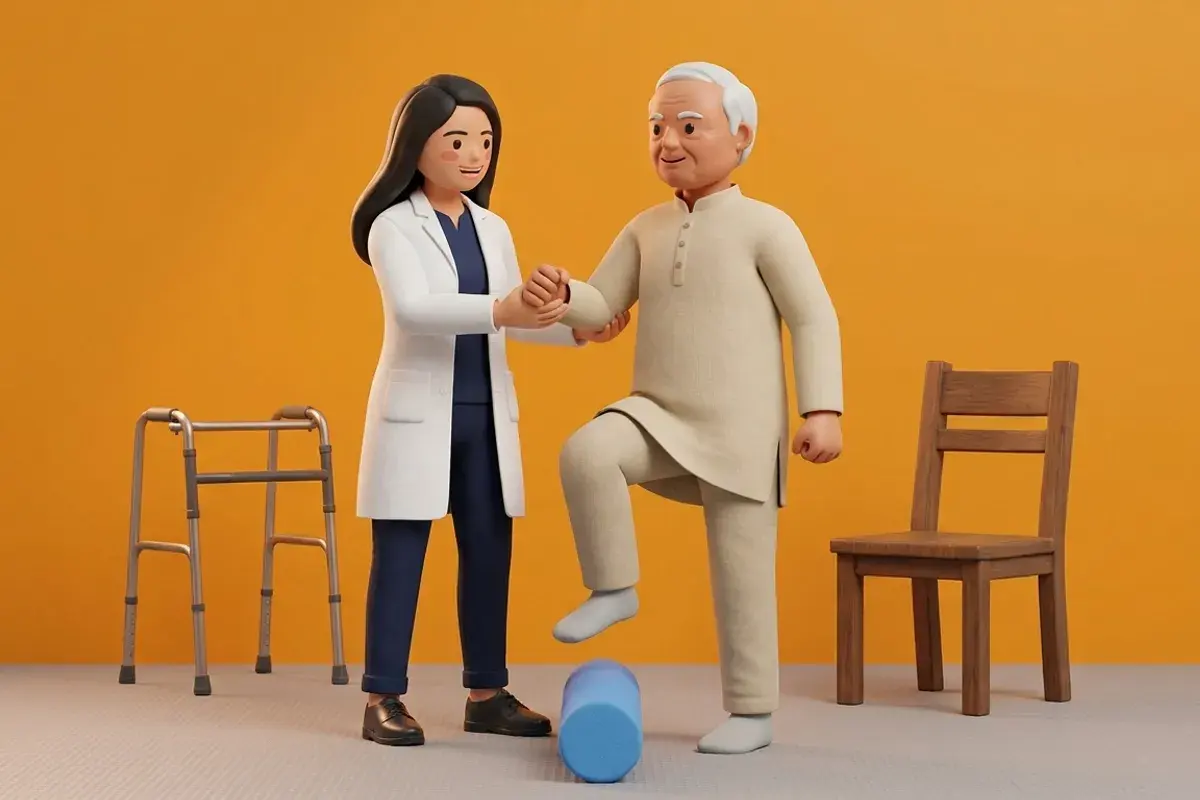
Touch-Down Weight Bearing (TDWB), also known as flat-foot contact, permits the foot of the operated leg to rest lightly on the floor for balance only, with vertical force limited to 10 to 15 kg (approximately the weight of the limb). Physiotherapists calibrate this tactile sensation using standard bathroom scales: the patient steps onto the scale until the needle reaches 10 to 12 kg, learning to reproduce this minimal pressure during 3-point crutch gait.
Prone Active-Assisted Hip Extension and Gluteal Firing
Objective: Reactivate the gluteus maximus without imparting compressive force across the anterior or superior acetabular dome.
- 1Assist the patient into a comfortable prone position with a folded pillow beneath the lower abdomen.
- 2Ensure the operated hip remains in neutral alignment without external rotation or abduction.
- 3Instruct the patient to gently tighten the lower abdominal muscles to stabilize the pelvis against the bed.
- 4Cue a smooth contraction of the gluteal muscle on the operated side, lifting the knee 1 to 2 cm off the bed.
- 5Hold the elevated contraction for three seconds while breathing out, then lower with control.
In patients operated for posterior wall or column acetabular fractures, never combine hip flexion beyond 80 degrees with internal rotation and adduction during the first two months. This combination creates direct posterior femoral head subluxation force.
Long-Term Surveillance and Complication Prevention
Ongoing surveillance focuses on detecting early post-traumatic osteoarthritis, heterotopic ossification, and avascular necrosis (AVN) of the femoral head. Periodic Judet oblique radiographs ensure long-term articular joint preservation.
- Femoral Head Sphericity Auditing: Review follow-up pelvic radiographs at 3, 6, and 12 months for subtle subchondral sclerosis or flattening of the femoral head dome.
- Trendelenburg Abductor Assessment: Evaluate gluteus medius strength dynamically during single-leg stance once full weight bearing is established, screening for superior gluteal nerve palsy.
- Sciatic Nerve Motor and Sensory Tracking: Continuously monitor common peroneal nerve sensation (dorsal foot) and active ankle dorsiflexion, common sites of traction injury following posterior surgical approaches.
Frequently Asked Questions
What is touch-down weight bearing and how is it different from partial weight bearing?
Touch-down weight bearing allows the foot to touch the floor purely for balance, bearing only 10 to 15 kg of weight (essentially limb weight). Partial weight bearing permits active loading of 25 to 50 percent of total body weight, requiring significantly more advanced bone consolidation.
Why are Judet radiographic views required before advancing my walking?
Standard front-facing (AP) X-rays do not show the three-dimensional curves of the acetabulum clearly. Judet views (iliac oblique and obturator oblique angles) reveal the anterior and posterior columns and the articular roof, confirming bone healing before weight is increased.
Do I need a doctor referral to begin home physiotherapy after complex trauma or external fixation?
Under the National Commission for Allied and Healthcare Professions (NCAHP) Act 2021, patients in India enjoy direct access to registered physiotherapists without requiring a mandatory doctor referral. At BookPhysio.in, you connect with verified clinical physiotherapists and pay directly per session without advance packages, hidden platform fees, or long-term contracts.
Related condition guides



Related recovery guides & articles
BookPhysio.in Editorial Team
The BookPhysio.in editorial team writes and maintains this content, checking it against reliable clinical sources. Content follows our medical review policy: general information only, and no page claims a clinical review until a named registered physiotherapist has signed it off.
Read our medical review policy