
Arthrofibrosis Prevention and Manipulation Under Anesthesia (MUA) Triage in Knee Arthroplasty
In short
Recognize the warning signs of arthrofibrosis early after knee arthroplasty, and understand the critical clinical window for manipulation under anesthesia (MUA).
In this guide (4 sections)
Arthrofibrosis, defined as an excessive, pathological fibroblastic proliferation and dense collagen deposition within the joint capsule, represents one of the most frustrating complications following total knee arthroplasty, affecting approximately five to ten percent of patients. Patients with arthrofibrosis experience progressive joint stiffness, loss of terminal extension, and failure to achieve functional flexion past 90 degrees, despite undergoing conventional rehabilitation. The condition transforms what should be a compliant, mobile joint into a rigid, fibrotic block encased in dense scar tissue. While aggressive prevention through early passive extension and active-assisted flexion is the primary defense, clinicians must recognize when conservative therapy has failed. When severe stiffness persists at six to eight weeks post-surgery, manipulation under anesthesia (MUA) offers an effective intervention to mechanically disrupt adhesions before permanent, mature collagen cross-links establish an irreversible contracture.
Stoicism, Fear of Exercise Pain, and Delayed Clinic Reviews in India
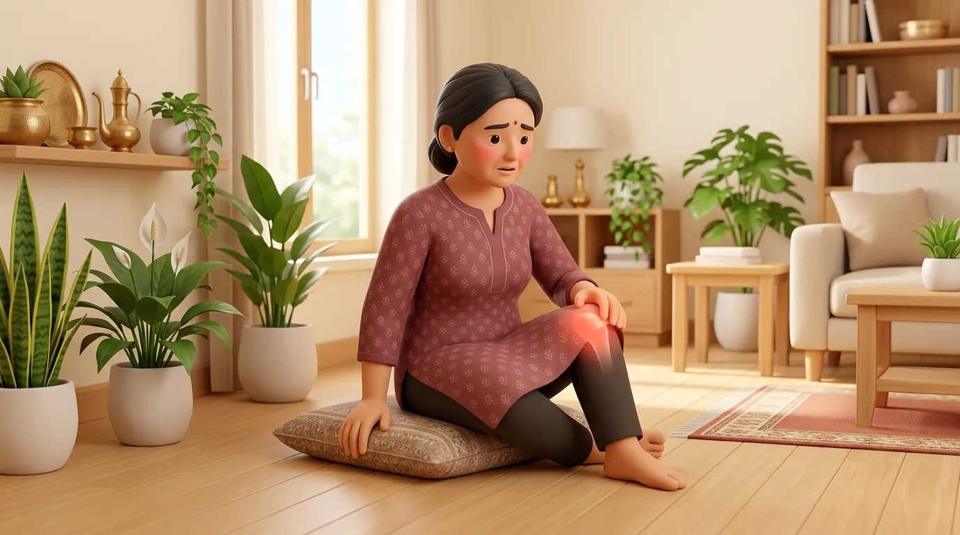
In Indian cultural contexts, two contrasting behavioral patterns often fuel arthrofibrosis development. In some households, elder patients exhibit extreme stoicism, quietly enduring joint stiffness without informing their family or attending scheduled outpatient physiotherapy sessions. In other cases, intense kinesiophobia (fear of movement pain) causes patients to refuse active knee bending, resting the limb in a semi-flexed position for weeks. Compounding these issues, patients residing in rural or semi-urban regions frequently delay returning to the operating hospital for their six-week follow-up due to transit logistics. Families must be educated that joint stiffness does not resolve on its own with time; missing the early clinical window allows soft tissue adhesions to solidify, making non-surgical recovery significantly harder.
Myofibroblast Transformation, Type I Collagen Shifts, and Adhesion Distribution
The cellular pathophysiology of arthrofibrosis is driven by an aberrant, prolonged inflammatory healing response. Following surgical tissue trauma, elevated levels of transforming growth factor-beta (TGF-β), platelet-derived growth factor (PDGF), and connective tissue growth factor (CTGF) stimulate normal fibroblasts to differentiate into contractile myofibroblasts expressing alpha-smooth muscle actin. Instead of concluding the normal wound healing cycle, these myofibroblasts synthesize excessive amounts of Type I and Type III collagen, depositing dense fibrotic sheets across the suprapatellar pouch, the medial and lateral gutters, and the intercondylar notch. As these collagen bundles cross-link into mature covalent bonds, they mechanically tether the extensor mechanism to the distal anterior femur. If closed manipulation is attempted after twelve weeks, the tensile strength of the mature scar tissue often exceeds that of host osteopenic bone, risking periprosthetic femoral fracture or patellar tendon avulsion rather than clean adhesion release.
Low-Load Long-Duration Stretching, Objective Goniometry, and MUA Timing
Preventing arthrofibrosis requires disciplined goniometric tracking and low-load, long-duration (LLLD) stretching protocols. Objective clinical red flags for impending arthrofibrosis include: failure to achieve 70 degrees of flexion by day fourteen, failure to achieve 90 degrees by week four, or a persistent extension deficit greater than 10 degrees at week six. Conservative management utilizes prolonged 15-minute passive traction holds rather than painful short-duration bounces. If goniometric range plateaus between weeks six and eight despite intensive therapy, urgent referral for MUA is executed. The ideal window for MUA is strictly between six and ten weeks post-surgery, where gentle closed manual flexion under regional anesthesia safely pops adhesions with minimal complication risk.
Low-Load Long-Duration (LLLD) Prone Hang Extension Stretch
Objective: Apply gentle, continuous gravitational elongation to remodel stubborn posterior capsular contractures
- 1Lie face down (prone) on a firm bed or treatment table with your lower legs extending out past the bottom edge.
- 2Position your thighs so your kneecaps rest just over the edge of the mattress, completely unsupported.
- 3Relax your thigh, hamstring, and gluteal muscles completely, allowing gravity to gently draw your lower legs toward the floor.
- 4To enhance elongation, loop a light 1-kilogram sandbag or soft strap around your operative ankle if tolerated without pain.
- 5Maintain this prolonged passive position for 10 to 12 minutes, taking slow deep breaths to prevent muscular guarding.
| Staging Classification | Post-Op Timing & Goniometric Range | Fibrotic Tissue Morphology | Recommended Clinical Protocol |
|---|---|---|---|
| Stage 1: Impending Arthrofibrosis | Week 2 to 4; flexion <80°, extension lag >10°; persistent joint warmth and tense effusion | Immature fibrin exudate; early myofibroblast migration into gutters | Intensify active-assisted ROM; LLLD stretching; cryotherapy; manual lymphatic drainage |
| Stage 2: Established Arthrofibrosis | Week 6 to 10; flexion stuck <90° despite compliant therapy; hard capsular end-feel | Organized Type III collagen bands in suprapatellar pouch and retinaculum | Urgent surgical consultation; schedule closed manipulation under anesthesia (MUA) |
| Stage 3: Chronic Mature Arthrofibrosis | Week >12; rigid contracture; flexion <85°; severe functional disability | Dense, cross-linked Type I collagen scar; bone remodeling; scar contracture | MUA contraindicated due to fracture risk; open or arthroscopic surgical lysis of adhesions |
Never permit a clinician to forcefully bounce or push your stiff knee with extreme body weight in an outpatient clinic setting without anesthesia. Violent manual force on an awake patient triggers maximal muscular resistance that can cause patellar tendon rupture or femoral condyle fracture. If closed manipulation is necessary, it must be performed safely in an operating theater under regional anesthesia.
Frequently Asked Questions
What happens during a manipulation under anesthesia (MUA), and does it damage my implant?
During an MUA, you are given light anesthesia or a regional block so your muscles are completely relaxed and you feel zero pain. The orthopedic surgeon gently and smoothly bends your knee through a full arc, listening and feeling for the soft tissue adhesions popping and releasing. The metal and plastic implants are completely undamaged. You wake up with immediate, painless range gains that must then be maintained with active physiotherapy.
Do I need a doctor referral for post-knee replacement physiotherapy on BookPhysio.in?
BookPhysio.in does not require a doctor referral. Under the National Commission for Allied and Healthcare Professions (NCAHP) Act 2021, patients across India enjoy direct access to autonomous registered physiotherapists. Through BookPhysio.in, you connect directly with verified clinicians and pay per session with transparent fees, completely free from advance package commitments, commission markups, or platform subscription charges.
Related condition guides



Related recovery guides & articles
BookPhysio.in Editorial Team
The BookPhysio.in editorial team writes and maintains this content, checking it against reliable clinical sources. Content follows our medical review policy: general information only, and no page claims a clinical review until a named registered physiotherapist has signed it off.
Read our medical review policy