
Arthroscopic Ankle Debridement for Anterolateral Impingement: Post-Surgical Rehab Protocols
In short
Explore the diagnosis, arthroscopic resection mechanics, and rapid functional rehabilitation protocol for chronic anterolateral soft-tissue impingement.
In this guide (4 sections)
Chronic anterolateral ankle impingement represents one of the most common long-term sequelae following an acute inversion sprain, accounting for persistent pain and disability in up to five percent of injured individuals. While the primary ligament tear (ATFL) may heal structurally, the accompanying intra-articular microtrauma and capsular tearing frequently trigger an aberrant, chronic inflammatory cascade within the anterolateral gutter of the talocrural joint. Repetitive micro-motion and joint instability induce marked synovial hyperplasia, fibrocartilaginous scar formation, and thickening of the accessory distal fascicle of the anterior inferior tibiofibular ligament (Bassett's ligament). When the ankle dorsiflexes, this hypertrophic fibrous mass becomes mechanically trapped between the anterior talus and the lateral malleolus, producing sharp, focal pain, localized swelling, and joint blocking. When conservative therapy fails, arthroscopic debridement delivers excellent outcomes, requiring a specialized post-operative rehabilitation protocol to restore joint function.
Navigating High Kerbs, Uneven Cobblestones, and Indian City Commutes
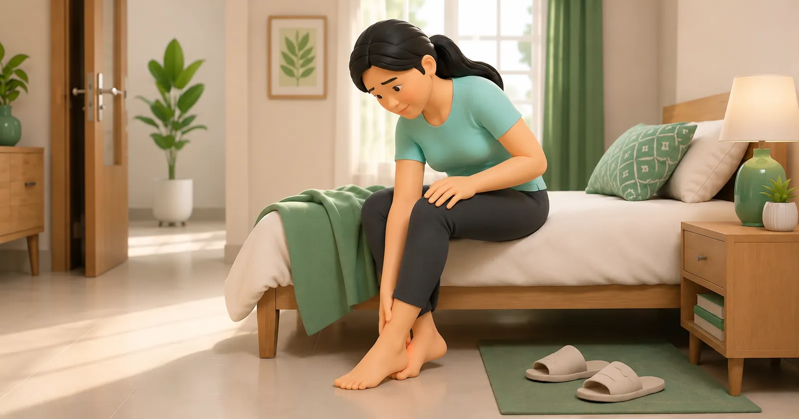
In Indian urban centers, living with chronic anterolateral impingement imposes substantial functional limitations. Walking along cracked footpaths or stepping down from high roadside concrete kerbs requires continuous closed-chain ankle dorsiflexion. Every time the patient steps down, the forward excursion of the tibia pinches the inflamed scar tissue in the anterolateral gutter, producing a sharp, disabling stab of pain. Commuting on public buses or suburban trains becomes intensely painful, as standing in swaying carriages forces the ankle into unexpected dorsiflexion-eversion postures that crush the sensitive synovial mass. Furthermore, ascending and descending steep staircases in apartment buildings without elevators triggers persistent perimalleolar aching and joint puffiness, forcing patients to avoid social and professional activities until the mechanical obstruction is surgically cleared and rehabilitated.
The Meniscoid Lesion, Bassett's Ligament Hypertrophy, and Arthrokinematic Jamming
The pathomorphology of anterolateral impingement centers on the formation of a distinct fibrocartilaginous mass classically described as a meniscoid lesion (Wolin's nodule). Following an inversion sprain, torn fibers of the anterior capsule and ATFL undergo repetitive mechanical trauma, stimulating local fibroblasts to synthesize a dense, disorganized collagenous scar covered with hypervascular synovium. Furthermore, the distal fascicle of the anterior inferior tibiofibular ligament (Bassett's ligament) often undergoes reactive hypertrophy. During normal ankle kinematics, Bassett's ligament clears the talar dome; however, when scarred or thickened, the anterolateral corner of the talar trochlea collides directly with this ligament during ankle dorsiflexion, shearing articular cartilage and producing chronic localized synovitis. During minimally invasive ankle arthroscopy, the surgeon utilizes small anteromedial and anterolateral portals to resect this hypertrophic tissue, restore gutter clearance, and ablate inflamed synovial fringes.
Rapid Mobilisation Protocol: Day One Weight-Bearing and Gutter Desensitisation
Unlike ligament reconstruction surgeries that require prolonged immobilization, post-arthroscopic debridement rehabilitation prioritizes early, aggressive functional mobilization. Because no structural ligamentous repair must be protected, patients are encouraged to bear weight as tolerated in a supportive sneaker on post-operative day one, using crutches only for two to three days for comfort. Registered physiotherapists implement immediate active range-of-motion exercises, emphasizing full closed-chain dorsiflexion to prevent the recurrence of fibrous adhesions within the newly debrided gutter. Manual therapy includes gentle cross-friction mobilization over portal scars and Maitland Grade III posterior talar glides. Within two to three weeks, patients progress to high-intensity stationary cycling, calf strengthening, dynamic balance board drills, and low-impact agility exercises, with full sports clearance typically achieved between four and eight weeks post-surgery.
Active Open-Chain Talocrural Range-of-Motion and Gutter Clearance Pumps
Objective: Maintain synovial fluid circulation and prevent the recurrence of fibrous adhesions in the debrided anterolateral gutter
- 1Sit on a comfortable chair or bed with your operated leg elevated on a pillow, removing any external bandage wraps as permitted.
- 2Slowly pull your toes and ankle upward into full active dorsiflexion, pausing for 2 seconds at the top of the movement.
- 3Smoothly point your foot downward into comfortable plantarflexion, pausing for 2 seconds.
- 4Perform slow, gentle circular movements with your foot, tracing 10 complete circles in a clockwise direction and 10 circles counterclockwise.
- 5Finish by actively spreading your toes wide and contracting your calf muscles gently for 5 seconds to pump away post-operative fluid.
| Post-Operative Recovery Milestone | Weight-Bearing and Protection Status | Targeted Physiotherapy Interventions | Clinical Functional Benchmark |
|---|---|---|---|
| Phase 1: Days 0 to 7 | Weight-bearing as tolerated in supportive shoe; wean off crutches by day 3 | Cryotherapy compression, elevation, active ankle pumps, gentle walking | Ambulate independently with normal heel-to-toe gait; minimal swelling |
| Phase 2: Weeks 1 to 3 | Full unassisted weight-bearing; no external bracing required | Maitland posterior talar glides, stationary cycling, double-leg calf raises | Weight-bearing lunge test distance equal to uninjured contralateral side |
| Phase 3: Weeks 3 to 6 | Full daily functional activity; normal footwear without restriction | Single-leg wobble board balance, eccentric calf lowering, light jogging | Complete 20 single-leg calf raises; run 2 kilometres pain-free |
| Phase 4: Weeks 6 to 8 | Unrestricted athletic training and full competitive match participation | Agility T-Test, lateral cutting drills, plyometric bounding | Limb Symmetry Index greater than 90 percent; full physician sports clearance |
If a patient continues to experience deep, aching joint line pain, mechanical catching, or recurrent joint effusion after arthroscopic soft-tissue debridement, an unrecognized osteochondral defect (OCD) of the talar dome or subchondral bone stress fracture must be evaluated. Pre-operative and post-operative MRI evaluation must carefully rule out chondral loss before attributing all symptoms solely to soft-tissue impingement.
Frequently Asked Questions
How long is the recovery after arthroscopic ankle debridement surgery?
Recovery after arthroscopic debridement is remarkably fast because no bones or ligaments were cut or repaired. Most patients walk comfortably without crutches within three to five days, return to desk jobs within one week, resume light jogging by three to four weeks, and return to full sports participation within six to eight weeks under structured physiotherapy.
Do I need a doctor referral to see an ankle sprain physiotherapist on BookPhysio.in?
BookPhysio.in does not require a doctor referral. Under the National Commission for Allied and Healthcare Professions (NCAHP) Act 2021, patients across India enjoy direct access to autonomous registered physiotherapists. Through BookPhysio.in, you connect directly with verified clinicians and pay per session with transparent fees, completely free from advance package commitments, commission markups, or platform subscription charges.
Related condition guides



Related recovery guides & articles
BookPhysio.in Editorial Team
The BookPhysio.in editorial team writes and maintains this content, checking it against reliable clinical sources. Content follows our medical review policy: general information only, and no page claims a clinical review until a named registered physiotherapist has signed it off.
Read our medical review policy