
Baker Cyst Management in Rheumatoid Knee Synovitis: Preventing Popliteal Rupture
In short
Popliteal Baker cysts in rheumatoid arthritis arise from persistent knee synovitis and high intra-articular pressure. Learn safe joint decompression protocols, gentle mobility arcs, and pseudothrombophlebitis prevention.
In this guide (6 sections)
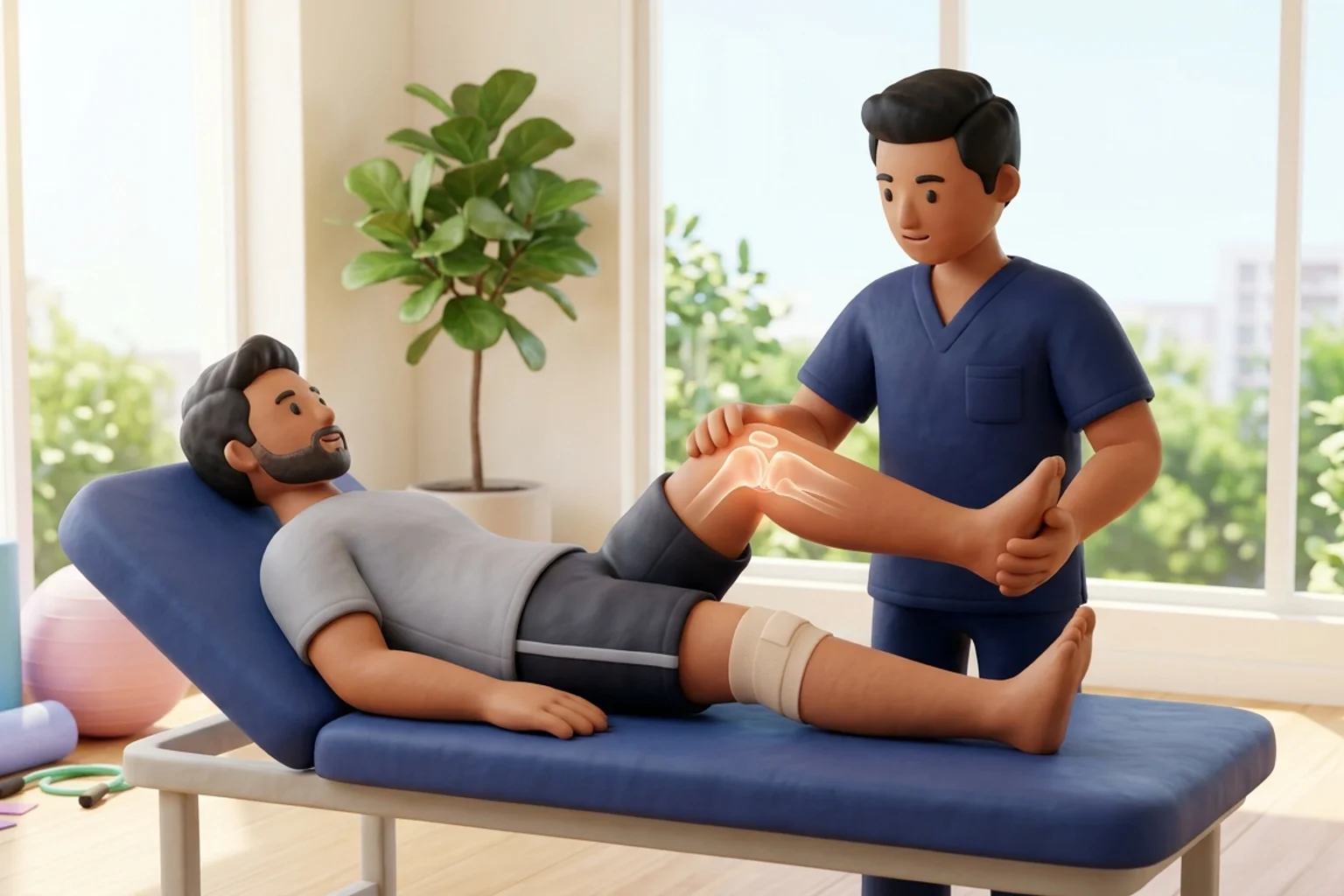
A popliteal cyst, commonly termed a Baker cyst, represents a distended, fluid-filled synovial bursa located in the posteromedial popliteal fossa, situated between the medial head of the gastrocnemius muscle and the semimembranosus tendon. In rheumatoid arthritis, chronic synovial inflammation produces excessive effusion, significantly elevating intra-articular hydrostatic pressure. A one-way valve-like communication between the knee joint capsule and the gastrocnemius-semimembranosus bursa permits synovial fluid to enter the cyst during knee flexion, but prevents its egress during extension. A massive or tense Baker cyst causes posterior knee fullness, limits terminal flexion, and risks spontaneous rupture. Popliteal rupture spills inflammatory synovial fluid into the deep calf fascial compartments, mimicking acute deep vein thrombosis (pseudothrombophlebitis).
Domestic and Cultural Indian Realities
In Indian domestic environments, daily habits often involve extreme knee flexion that dramatically elevates popliteal pressure. Squatting completely to use an Indian ground commode, sitting cross-legged on the floor (sukhasana) during meals or religious rituals, and kneeling to clean floor corners compress the popliteal space directly, spiking intra-articular pressure over 300 mmHg. In the presence of a tense Baker cyst, these high-pressure moments can trigger catastrophic cyst rupture into the calf. Furthermore, traditional masseurs often attempt to forcefully knead away the popliteal lump with deep thumb pressure. This practice is extremely hazardous and must be strictly prohibited. Patients must transition to high chairs and Western commodes, completely avoiding deep knee hyperflexion.
Clinical Biomechanics and Inflammatory Pathology
The formation and expansion of a Baker cyst is a secondary phenomenon driven by primary intra-articular knee pathology. In rheumatoid arthritis, extensive villous synovial hyperplasia and excessive exudate accumulation raise baseline resting joint pressure. During active knee extension, the anterior joint capsule tenses, forcing synovial fluid posteriorly. During deep flexion past 90 degrees, the semimembranosus and gastrocnemius muscles contract, compressing the transverse capsular slit and trapping fluid within the bursa. When the cyst wall ruptures under excessive hydraulic strain, pro-inflammatory fluid dissects along the soleus and gastrocnemius muscle planes, triggering acute chemical cellulitis and compartment oedema.
Clinical Assessment and Functional Staging
Clinical evaluation begins with Foucher sign: the popliteal cyst is palpated with the knee in full extension, where it feels firm, tense, and prominent, and then in 45 degrees of flexion, where it softens and becomes ballotable as capsular tension releases. Point-of-care musculoskeletal ultrasound is the gold standard diagnostic tool, confirming the anechoic cystic structure, its anatomical neck between the semimembranosus and gastrocnemius tendons, and ruling out popliteal artery aneurysm. In patients presenting with acute calf pain and swelling, emergency Doppler ultrasound is mandatory to differentiate ruptured Baker cyst from acute deep vein thrombosis.
Targeted Mobility and Joint Preservation Exercises
Physiotherapy must avoid aggressive end-range passive knee flexion, heavy resisted squats, and direct popliteal compression. Exercises emphasize non-weight bearing rhythmic motion to circulate fluid and gentle isometric quadriceps strengthening.
Seated Gentle Pendulum Knee Swings in Mid-Range Arc
Objective: Lower intra-articular hydrostatic pressure and promote synovial fluid circulation without compressing the popliteal space
- 1Sit on a high table or sturdy treatment surface so your lower legs hang freely without touching the floor.
- 2Gently swing your lower legs forward and backward in a smooth, rhythmic pendulum arc between 30 and 70 degrees of flexion.
- 3Do not force the knees into full extension or pull them tightly into deep flexion.
- 4Continue the gentle swinging motion rhythmically for 2 minutes while maintaining relaxed breathing.
| Intervention Domain | Joint-Preserving Practice | High-Rupture-Risk Practice |
|---|---|---|
| Knee Range of Motion | Gentle active-assisted movement confined to 20 to 80 degrees of comfortable flexion | Forced passive terminal knee flexion stretches, kneeling, or deep squatting |
| Manual Therapy | Light retrograde lymphatic drainage of the thigh and leg; zero pressure over popliteal fossa | Deep friction massage, vigorous kneading, or thumb compression over the popliteal cyst |
| Sanitation & Seating | Elevated Western toilet seat and dining chairs with seat height at 19 inches | Ground-level squatting commodes and cross-legged sitting on floor mats |
| Calf Symptoms | Immediate duplex Doppler ultrasound if calf swelling develops to rule out DVT | Applying heat packs or massaging an acutely swollen, painful calf |
Red Flags and Immediate Rheumatology Escalation
Sudden, severe, tearing calf pain accompanied by rapid leg swelling, warmth, and crescent-shaped bruising around the malleoli (crescent sign) signals an acute popliteal cyst rupture. Immediate emergency Doppler ultrasound is mandatory to exclude concurrent deep vein thrombosis, as the clinical presentation of pseudothrombophlebitis is indistinguishable from true venous thromboembolism without vascular imaging.
Frequently Asked Questions
Can physiotherapy exercises make a Baker cyst disappear completely?
A Baker cyst is driven by underlying knee joint synovitis. Physiotherapy alone cannot eliminate the cyst if active joint inflammation persists. However, by managing intra-articular pressures, preventing high-risk flexion postures, and strengthening knee musculature, physiotherapy helps decompress the cyst, relieves posterior knee tightness, and prevents popliteal rupture.
How do I book a specialised knee physiotherapy consultation at home through BookPhysio.in?
Under the National Commission for Allied and Healthcare Professions (NCAHP) Act 2021, Indian patients have direct access to registered physiotherapists without an obligatory physician referral. On BookPhysio.in, you schedule an appointment with a verified clinician in your city. Payment is made directly to the therapist at your home appointment via cash or UPI; BookPhysio.in does not collect booking fees online.
Related condition guides



Related recovery guides & articles
BookPhysio.in Editorial Team
The BookPhysio.in editorial team writes and maintains this content, checking it against reliable clinical sources. Content follows our medical review policy: general information only, and no page claims a clinical review until a named registered physiotherapist has signed it off.
Read our medical review policy