
Cardiac Telemetry and ECG Monitoring in Exercise: Arrhythmia Identification and Safety Cut-Offs
In short
Real-time ECG telemetry ensures patient safety during cardiac rehabilitation. Learn dangerous arrhythmia patterns, ST segment ischemic changes, and mandatory stop rules.

In this guide (5 sections)
The Critical Role of ECG Telemetry in Phase 2 Cardiac Rehab
During the early outpatient phase of cardiac rehabilitation (Phase 2), patients recovering from myocardial infarction, coronary stenting, or cardiac surgery possess an electrically unstable myocardium vulnerable to exercise-induced arrhythmias.
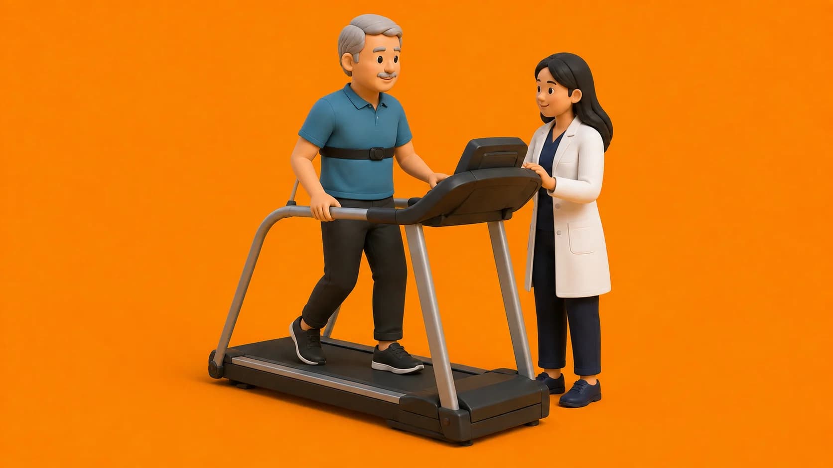
Continuous electrocardiographic (ECG) telemetry allows the cardiopulmonary physiotherapist to monitor heart rate, cardiac rhythm, and ST-segment displacement in real time as myocardial oxygen demand increases.
Risk stratification (AACVPR guidelines) categorizes patients into Low, Moderate, and High risk, determining the duration and necessity of continuous ECG monitoring.
Dangerous ECG Arrhythmias and Ischemic Signs During Exercise
Immediate clinical recognition of abnormal rhythm strips dictates whether exercise should be modified, paused, or terminated with emergency activation.
| ECG Manifestation | Rhythm Characteristics | Clinical Significance | Mandatory Action | |
|---|---|---|---|---|
| 1. Isolated Unifocal PVCs | Occasional premature ventricular complexes (< 6/min) | Benign ectopic focus; normal in deconditioned hearts | Continue exercise with close monitoring; record frequency. | |
| 2. Multifocal PVCs / Couplets | PVCs with varying morphology or two in a row | Indicates increasing myocardial irritability and ischemia | Reduce exercise workload; re-check blood pressure and SpO2. | |
| 3. Ventricular Tachycardia (VT) | Run of 3 or more consecutive wide-complex PVCs | Lethal hemodynamically unstable arrhythmia | Stop exercise immediately; emergency medical protocol; crash cart. | |
| 4. Horizontal ST Depression | >= 1 mm horizontal or downsloping ST depression | Signifies subendocardial myocardial ischemia | Stop exercise; place patient seated; check vitals and sublingual nitrate. | |
| 5. Second or Third Degree AV Block | Progressive PR prolongation (Mobitz I) or dropped beats | Impaired atrioventricular nodal conduction | Terminate exercise immediately; cardiology review for pacemaker. |
Absolute vs Relative Exercise Termination Cut-Offs
The American College of Sports Medicine (ACSM) and American Heart Association (AHA) establish clear termination criteria during clinical exercise training.
- Absolute Stop: Angina Pectoris: Onset of moderate to severe angina (Grade 2 or higher on 4-point angina scale) with retrosternal pressure or jaw/left arm radiation.
- Absolute Stop: Hemodynamic Collapse: Drop in systolic blood pressure of > 10 mmHg below baseline despite increasing workload, accompanied by signs of hypoperfusion (pallor, ataxia, dizziness).
- Absolute Stop: Hypertensive Crisis: Systolic blood pressure exceeding 250 mmHg or diastolic blood pressure exceeding 115 mmHg.
- Absolute Stop: Sustained Tachyarrhythmias: Sustained ventricular tachycardia, new-onset rapid atrial fibrillation (HR > 140 bpm), or multiple ventricular triplets.
Electrolyte Imbalances and Common Triggers in India
In Indian cardiac patients on potent loop diuretics (such as furosemide or torsemide) combined with ACE inhibitors, severe summer sweating frequently leads to hypokalemia (serum potassium < 3.5 mEq/L) or hypomagnesemia.
Electrolyte depletion drastically lowers ventricular fibrillation thresholds, triggering sudden bursts of ventricular ectopy during routine treadmill walks. Physiotherapists review recent electrolyte panels before initiating high workloads.
Frequently Asked Questions
What is the difference between upsloping and horizontal ST depression?
Rapid upsloping ST depression is often a normal physiological response to high heart rates in fit individuals. Horizontal or downsloping ST depression (>= 1 mm measured 80 ms past the J-point) indicates true myocardial ischemia and mandates exercise cessation.
How long must a cardiac patient wear telemetry during Phase 2 rehab?
Low-risk patients typically require telemetry for 6 to 12 sessions. Moderate to high-risk patients (those with low ejection fraction, complex arrhythmias, or previous cardiac arrest) may require continuous telemetry for 24 to 36 sessions.
Can exercise trigger atrial fibrillation?
Yes. Intense sympathetic stimulation or sudden blood pressure surges can trigger paroxysmal atrial fibrillation in predisposed patients. If the ventricular rate becomes rapid, exercise is stopped.
Related condition guides

Related recovery guides & articles
BookPhysio.in Editorial Team
The BookPhysio.in editorial team writes and maintains this content, checking it against reliable clinical sources. Content follows our medical review policy: general information only, and no page claims a clinical review until a named registered physiotherapist has signed it off.
Read our medical review policy