
Cuboid and Navicular Joint Glides: Mobilizing Midfoot Hypomobility in Plantar Fasciopathy
In short
Learn how cuboid and navicular joint mobilizations restore midfoot arthrokinematics, resolve cuboid syndrome, and relieve chronic tension on the plantar fascia.
In this guide (4 sections)
While orthopaedic evaluation of plantar heel pain routinely examines the calcaneus and ankle, the transverse tarsal (Chopart) joint complex, specifically the navicular and cuboid articulations, is frequently the neglected biomechanical epicenter of persistent foot dysfunction. The navicular bone serves as the mechanical keystone of the medial longitudinal arch, articulating with the talar head and the three cuneiforms, while the cuboid anchors the lateral longitudinal column, providing a dynamic pulley for the peroneus longus tendon. During normal gait, these midfoot bones must execute coordinated arthrokinematic glides: transitioning from a mobile, unlocked configuration during weight acceptance to absorb impact shock, into a rigid, close-packed locked configuration during terminal stance to facilitate propulsion. Hypomobility or subtle subluxation of the cuboid or navicular disrupts this delicate joint harmony, altering load distribution and transferring excessive, chronic tensile shear into the medial and central slips of the plantar aponeurosis.
Barefoot Marble Floor Living, Prolonged Kitchen Standing, and Thin Flat Slippers
In Indian households, midfoot joint hypomobility is exceptionally common due to cultural footwear and domestic flooring conditions. The rigid, unyielding surface of polished marble and granite flooring offers no mechanical compliance. When individuals walk barefoot across these flat stone slabs, the midfoot joints are subjected to repetitive, unbuffered vertical impacts. Furthermore, wearing popular traditional footwear like stiff flat Kolhapuris or flimsy rubber chappals deprives the lateral and medial arches of proper anatomical support. In individuals who frequently navigate uneven stone footpaths or step off public buses onto broken curbs, subtle inversion-plantarflexion trauma can subtly subluxate the cuboid downward (cuboid syndrome) or fixate the navicular in a dropped position. When these individuals return home to cook barefoot or perform chores, the locked midfoot transmits relentless tensile strain directly into the calcaneal enthesis.
Windlass Mechanism Strain, Calcaneal Enthesopathy, and Plantar Thickness
The pathomechanics linking midfoot hypomobility to plantar fasciopathy operate through altered midtarsal joint locking mechanisms. When the navicular bone becomes fixated in a downward, pronated position (navicular drop), the talonavicular joint cannot resupinate during the late stance phase of gait. This failure to lock keeps the midfoot flexible when it needs to be rigid, forcing the central band of the plantar fascia to bear excessive tensile elongation loads that should normally be shared by interlocking tarsal bones. Conversely, a subluxated or hypomobile cuboid disrupts the mechanical pulley of the peroneus longus tendon, preventing it from depressing the first metatarsal head to stabilize the medial column. Ultrasound evaluations in these patients consistently demonstrate asymmetrical thickening of the proximal plantar fascia, accompanied by localized peritendinous inflammation and pain that resists standard calf stretches.
Rathleff High-Load Towel Training, Low-Dye Taping, and Intrinsic Foot Doming
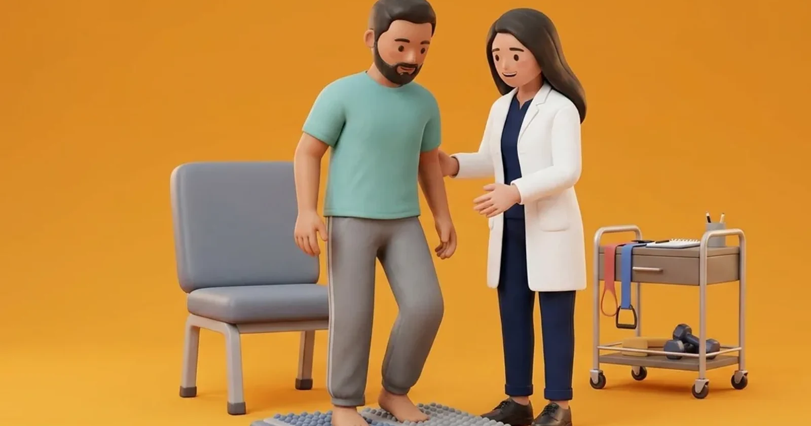
Restoring midfoot mobility requires targeted manual joint mobilization techniques delivered by an experienced physiotherapist. For navicular hypomobility, clinicians apply passive plantar-to-dorsal glides, using the thumbs to mobilize the navicular tuberosity upward while stabilizing the talus. For cuboid hypomobility or cuboid syndrome, the cuboid whip or plantar-dorsal glide restores normal lateral column arthrokinematics. Following mobilization, low-Dye taping or a supportive midfoot pad is applied immediately to maintain joint alignment during early walking. Once midfoot mobility is restored, patients transition into active rehabilitation: practicing the Rathleff high-load strength protocol, eccentric calf loading, and short-foot doming drills to train the intrinsic musculature and the tibialis posterior to maintain dynamic arch integrity.
Tennis Ball Seated Midfoot Arch Mobilization and Rolling
Objective: Gently mobilize hypomobile tarsal joints and relieve deep intrinsic fascial tension across the midfoot
- 1Sit comfortably in a chair with your bare foot placed over a firm tennis ball or massage ball resting on a non-slip floor mat.
- 2Position the ball directly beneath the middle of your foot arch, between your heel and the ball of your foot.
- 3Apply light to moderate downward pressure, slowly rolling the ball in small circular motions directly under the navicular and cuboid bones.
- 4Pause on tight, stiff areas for 10 to 15 seconds, taking slow, deep diaphragmatic breaths while allowing your foot muscles to relax over the ball.
- 5Continue for 2 to 3 minutes, avoiding aggressive, painful pressure directly over the heel bone.
| Articular Structure | Manual Mobilization Technique | Clinical Indication & Signs | Biomechanical Impact on Plantar Fascia |
|---|---|---|---|
| Navicular Plantar-to-Dorsal Glide | Bimanual thumbs apply upward dorsal pressure to navicular tuberosity with talus fixed | Navicular fixed in pronated drop; restricted midfoot supination at push-off | Restores medial arch height and relieves excessive tensile stretch on the fascia |
| Cuboid Plantar-to-Dorsal Glide (Whip) | Thumbs placed on plantar aspect of cuboid; gentle dorsal thrust with forefoot traction | Cuboid syndrome; lateral foot ache, restricted eversion, feeling of walking on a pebble | Restores peroneus longus pulley action, stabilizing the 1st ray and medial column |
| Cuneometatarsal Joint Glides | Planar dorsal and plantar glides of individual metatarsal bases on cuneiforms | Transverse arch rigidity; stiff, flat forefoot with lack of adaptive splay | Expands forefoot ground contact area and dampens shock transmitted to the calcaneus |
Carefully differentiate midfoot joint stiffness from midtarsal stress fractures, particularly navicular stress fractures which present with focal bone tenderness over the 'N-spot' (dorsal navicular crest). If severe, deep bone pain is reproduced by navicular palpation or calcaneal squeezing, immediately discontinue joint mobilizations. Obtain prompt orthopaedic consultation and urgent MRI.
Frequently Asked Questions
What is cuboid syndrome, and can it cause pain that feels like plantar fasciitis?
Cuboid syndrome occurs when the cuboid bone, located on the outer edge of your midfoot, subtly shifts or becomes stuck out of alignment, usually following an ankle sprain or from walking on hard, uneven surfaces. Because the cuboid helps anchor the foot muscles and ligaments, a stuck cuboid causes deep aching across your arch and heel that closely mimics plantar fasciitis. A skilled physiotherapist can perform a gentle manual adjustment (cuboid glide) to restore the bone's alignment, providing immediate relief.
Do I need a doctor referral to treat plantar fasciitis on BookPhysio.in?
BookPhysio.in does not require a doctor referral. Under the National Commission for Allied and Healthcare Professions (NCAHP) Act 2021, patients across India enjoy direct access to autonomous registered physiotherapists. Through BookPhysio.in, you connect directly with verified clinicians and pay per session with transparent fees, completely free from advance package commitments, commission markups, or platform subscription charges.
Related condition guides



Related recovery guides & articles
BookPhysio.in Editorial Team
The BookPhysio.in editorial team writes and maintains this content, checking it against reliable clinical sources. Content follows our medical review policy: general information only, and no page claims a clinical review until a named registered physiotherapist has signed it off.
Read our medical review policy