
Day Zero to Week Two: ACL Graft Protection and Early Patellar Mobilization
In short
Master the critical first two weeks post-ACL reconstruction: managing surgical effusion, achieving full passive extension, and preventing soft tissue adhesions.
In this guide (4 sections)
The first fourteen days following ACL reconstruction represent a critical biological window where the foundation of long-term knee function is established. During this immediate post-operative phase, the surgically anchored graft is at its strongest mechanical baseline because fixation depends entirely on hardware: titanium or bioabsorbable interference screws, adjustable cortical loop buttons, or cross-pin anchors. However, the surrounding knee tissues are traumatized by surgical incisions, portal drilling, and intra-articular debris, initiating a vigorous acute inflammatory cascade. The primary clinical objectives during this phase are strictly protective: controlling surgical pain and hemarthrosis, safeguarding graft tunnel fixation, preventing deep vein thrombosis (DVT), restoring complete passive knee extension matching the contralateral side, and preventing patellofemoral adhesions through meticulous patellar mobilization.
Navigating Western vs Indian Toilets, Bathroom Slipping, and Bed Transfers
In Indian households, the immediate post-operative period introduces immediate environmental safety challenges. Wet bathroom floors, common in Indian homes where bucket baths or wet-area washrooms are standard, present a severe slipping hazard for a patient wearing an immobilizer brace and navigating with axillary crutches. Patients must use anti-skid rubber footwear and dry towels across floor surfaces. Traditional squat toilets are completely contraindicated because deep knee flexion will tear fresh surgical repairs; homes with only Indian commodes must install a temporary raised toilet chair with armrests. Bed transfers must be executed smoothly without letting the surgical leg drag or hang unsupported, which places sudden rotational shear on fresh tunnel fixations.
Biological Inflammatory Phase, Fibroblast Ingrowth, and Retinacular Adhesions
Histologically, days 0 to 14 correspond to the acute necrosis and inflammatory phase of graft incorporation. Within hours of tourniquet release, a fibrin clot envelops the intra-articular graft segment and fills the bone tunnels. Neutrophils and macrophages infiltrate the graft substance to clear cellular debris, releasing cytokines that signal intrinsic tenocytes and extrinsic synovial fibroblasts to initiate collagen synthesis. Concurrently, surgical trauma to the suprapatellar pouch and infrapatellar fat pad triggers rapid fibrin deposition. If the patella is allowed to remain motionless, dense fibrous bands adhere the patella to the anterior femoral trochlea and anterior tibial cortex, producing a stiff, painful extensor mechanism that resists future flexion and terminal extension.
Acute Protection Protocol: Cryotherapy, Patellar Glides, and Ankle Pump Cycles
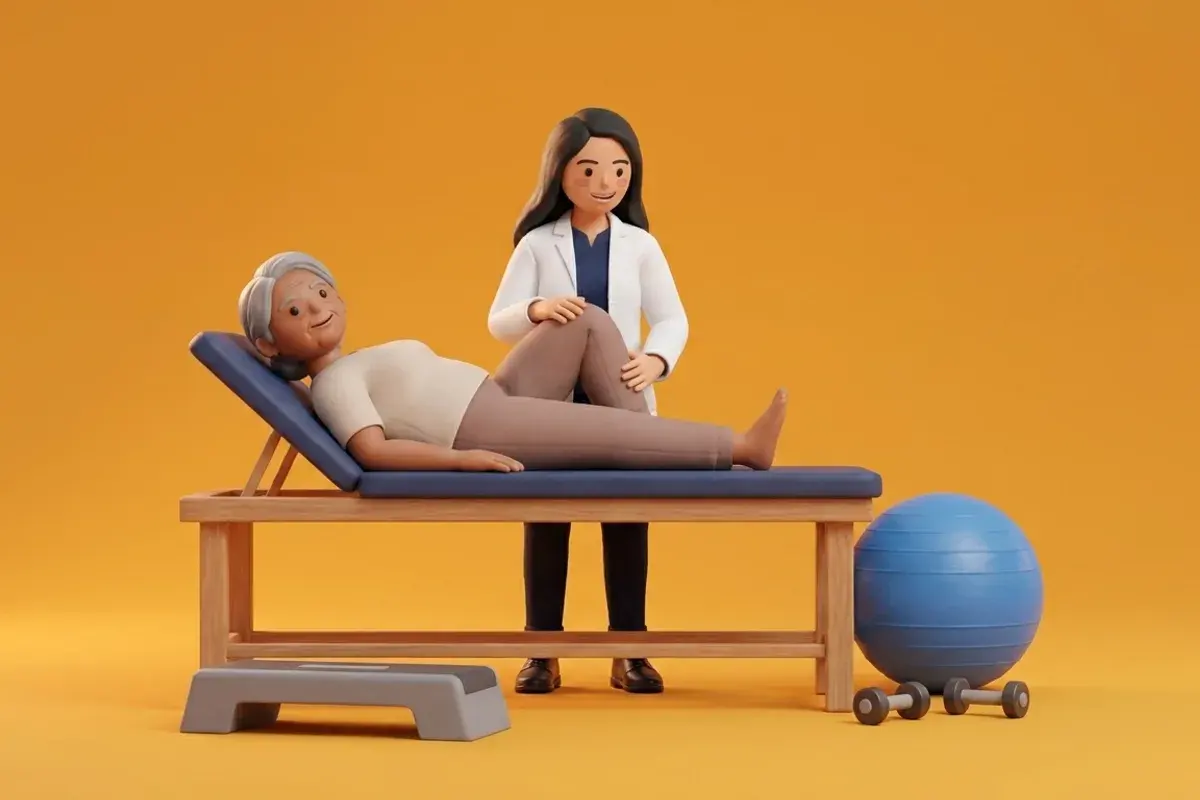
The early rehabilitation prescription combines total graft protection with active mobilization of non-compromised adjacent tissues. Cryo-compression therapy applied for 20 minutes every 2 hours suppresses local tissue temperature, reducing vascular permeability and metabolic demand. Multi-directional patellar mobilizations (superior, inferior, medial, lateral) must be performed multiple times daily with the quadriceps completely relaxed. Ankle pump routines (30 repetitions every waking hour) stimulate the calf muscle venous pump to prevent venous stasis and thromboembolism. Controlled weight bearing with crutches and a locked extension brace is commenced according to surgical guidelines, ensuring the patient walks with a heel-to-toe gait without limping.
Active Ankle Dorsiflexion-Plantarflexion Venous Pumps
Objective: Stimulate the deep calf venous muscle pump to prevent lower extremity venous stasis and deep vein thrombosis
- 1Lie comfortably in bed with your surgical leg elevated on two pillows positioned lengthwise under the calf and foot.
- 2Keep your knee in its locked straight extension brace.
- 3Pull your toes and foot firmly upward toward your shin (dorsiflexion) as far as comfortably possible, holding for 1 second.
- 4Point your toes and foot downward away from your body (plantarflexion), squeezing your calf muscle firmly for 1 second.
- 5Repeat these rhythmic pumping movements continuously and smoothly without rotating your hip.
| Evaluation Metric | Target Milestone (Day 7) | Target Milestone (Day 14) | Intervention if Target Missed |
|---|---|---|---|
| Passive Knee Extension | 0 degrees (neutral extension) | Full hyperextension matching contralateral knee | Initiate low-load prolonged stretch with prone heel hangs and towel props |
| Active Knee Flexion | 70 to 80 degrees | 90 degrees comfortably without force | Gentle assisted heel slides on smooth board with towel strap assistance |
| Quadriceps Activation | Flicker contraction of VMO visible | Straight leg raise performed without extensor lag | Implement high-intensity NMES at 50 Hz with cryotherapy pre-treatment |
| Wound and Effusion Status | Clean, dry portals with dressing intact | Sutures/staples removed, trace to 1+ effusion | Elevate limb continuously, enforce strict cryo-compression, verify portal healing |
Asymmetric swelling of the lower leg, increasing persistent dull ache in the calf belly, skin warmth, and localized tenderness upon calf palpation are clinical hallmarks of deep vein thrombosis (DVT). If accompanied by sudden shortness of breath or pleuritic chest pain, treat as an emergency pulmonary embolism. Seek urgent vascular Doppler evaluation and immediate emergency care.
Frequently Asked Questions
Should I sleep with a pillow under my operated knee to reduce post-operative pain?
No, placing a pillow directly underneath the knee is one of the most detrimental mistakes during early recovery. While a bent knee feels temporarily comfortable by reducing capsular tension, resting in flexion encourages the hamstring muscles and posterior capsule to tighten. This rapidly causes a permanent knee extension deficit that is exceptionally difficult to correct later. Always place pillows under the ankle or calf, keeping the knee straight.
Do I need a doctor referral to rehabilitate an ACL reconstruction on BookPhysio.in?
BookPhysio.in does not require a doctor referral. Under the National Commission for Allied and Healthcare Professions (NCAHP) Act 2021, patients across India enjoy direct access to autonomous registered physiotherapists. Through BookPhysio.in, you connect directly with verified clinicians and pay per session with transparent fees, completely free from advance package commitments, commission markups, or platform subscription charges.
Related condition guides



Related recovery guides & articles
BookPhysio.in Editorial Team
The BookPhysio.in editorial team writes and maintains this content, checking it against reliable clinical sources. Content follows our medical review policy: general information only, and no page claims a clinical review until a named registered physiotherapist has signed it off.
Read our medical review policy