
Delayed Union and Non-Union Triage: Persistent Focal Tenderness Post-Fracture
In short
Recognize the clinical warning signs of delayed union and non-union, manage persistent bone tenderness, and navigate orthopaedic revision triage.
In this guide (4 sections)
While the vast majority of ankle fractures heal uneventfully following appropriate anatomical reduction and stable internal fixation, a significant minority of patients experience impaired bone healing. In clinical orthopaedics, delayed union is defined as the failure of a fracture to achieve solid osseous consolidation within three to six months following injury, whereas non-union represents the complete cessation of reparative osteogenesis without bone union after six to nine months. Identifying impaired healing early in the rehabilitation journey is essential to prevent permanent disability, catastrophic hardware fatigue failure, or progressive joint malalignment. Physiotherapists occupying primary-contact roles must maintain high diagnostic vigilance, recognizing the subtle clinical, biomechanical, and radiographic signs that distinguish expected post-surgical discomfort from biological bone non-union.
Coping with Lingering Bone Pain During Daily Errands and Domestic Tasks
In Indian households, an individual struggling with delayed bone union faces compounding domestic frustrations. Months after friends and family expect them to be fully recovered, the patient still experiences deep, throbbing bone pain whenever attempting to walk to the local grocery shop or stand at the kitchen counter to prepare meals. Stepping onto uneven domestic tiles, climbing steep residential staircases, or negotiating broken street kerbs triggers sharp, sickening pain directly over the fibular plate or medial malleolar screws. Family members often question whether the pain is psychological or due to lack of effort, creating emotional distress. The patient frequently notices that their evening swelling worsens rather than improves over time, forcing them back onto crutches. Clinical validation and objective diagnostic triage are essential to guide the patient toward appropriate revision care.
Hypertrophic Versus Atrophic Non-Union, Strain Theory, and Hardware Fatigue
The pathomechanics of impaired bone healing reflect a failure of either biology, mechanics, or both. Non-unions are categorized broadly into hypertrophic (vascular, biologically active, but mechanically unstable, displaying elephant-foot callus without bridging) and atrophic (biologically inert, avascular, displaying rounded, osteopenic bone ends without callus formation). According to Perren's tissue strain theory, bone formation can occur only if local interfragmentary strain remains below two percent. If rigid fixation is lost or excessive early axial loading causes persistent micro-motion exceeding ten percent, osteoblasts cannot bridge the gap, and fibrous non-union develops. Concurrently, cyclical mechanical loading is transferred entirely to the metallic plate and screws; because titanium and stainless steel have finite fatigue life thresholds, persistent non-union inevitably culminates in implant fatigue failure and hardware snapping.
Clinical Triage Protocol: Palpation Tenderness, Tuning Fork Tests, and Unloading
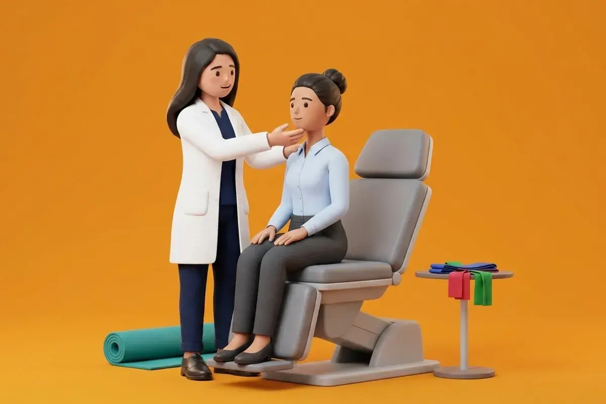
Physiotherapists identify delayed union through a multi-modal assessment battery. Point tenderness localized precisely over the fracture line upon direct thumb pressure represents the single most sensitive clinical marker of incomplete osseous union. Clinicians perform an acoustic tuning fork test: placing a vibrating one-hundred-and-twenty-eight-Hertz tuning fork directly over the fracture line elicits a sharp, deep bone ache if non-union exists, whereas healed bone transmits vibrations painlessly. When delayed union is suspected, aggressive weight-bearing and manual joint mobilizations are suspended immediately. Treatment incorporates biophysical stimulation modalities, such as Low-Intensity Pulsed Ultrasound (LIPUS) or capacitive coupling bone growth stimulators, while transitioning the patient into a rigid pneumatic walking boot. Prompt orthopaedic consultation is arranged to evaluate advanced computed tomography (CT) scans and plan potential autologous bone grafting or revision plating.
Non-Weight-Bearing Towel-Assisted Isometric Ankle Stabilization
Objective: Stimulate lower leg muscle activation and local blood circulation without transmitting micro-motion across an ununited fracture
- 1Sit comfortably in a supportive armchair with your recovering leg elevated horizontally on a soft ottoman or bed.
- 2Loop a non-elastic cotton towel or dupatta around the ball of your foot, holding the ends securely with both hands.
- 3Gently press the ball of your foot forward into the towel as if lightly pressing a car accelerator, using only twenty-five percent effort.
- 4Hold the towel firmly so that your ankle remains completely motionless, producing a purely static, isometric contraction in your calf.
- 5Maintain the gentle contraction for six seconds, breathe calmly, and then relax completely for four seconds before repeating.
| Non-Union Classification | Biological & Radiographic Appearance | Primary Etiological Mechanism | Targeted Orthopaedic & Physical Intervention |
|---|---|---|---|
| Delayed Union (3-6 Months) | Faint fracture line remains; minimal bridging callus | Inadequate mechanical stability or impaired vascularity | Pneumatic boot offloading; LIPUS ultrasound; protected PWB |
| Hypertrophic Non-Union (> 6 Mos) | Abundant non-bridging callus (elephant foot appearance) | Mechanical instability with preserved biological blood supply | Revision rigid internal fixation with compression plating |
| Atrophic Non-Union (> 6 Mos) | Rounded, sclerotic bone ends; zero callus formation | Biological failure; avascularity; infection; smoking | Autologous bone graft harvest; biological revision ORIF |
If a patient with suspected delayed union experiences an acute, audible 'crack' or sudden sharp snap during walking, followed by an immediate return of instability and localized swelling, metallic hardware fatigue failure has occurred. Cease all weight-bearing immediately, immobilize the limb in a rigid splint, and arrange for immediate orthopaedic evaluation and emergency radiographs.
Frequently Asked Questions
How can I tell if my ankle bone is not healing properly?
The clearest warning sign of poor bone healing is persistent, sharp pain directly over the bone whenever you touch it or put weight on it, several months after your surgery. If your ankle continues to throb every evening, feels loose, or hurts just as much at three months as it did at six weeks, your physiotherapist will refer you for a CT scan to check your bone healing.
Do I need a doctor referral to rehabilitate an ankle fracture on BookPhysio.in?
BookPhysio.in does not require a doctor referral. Under the National Commission for Allied and Healthcare Professions (NCAHP) Act 2021, patients across India enjoy direct access to autonomous registered physiotherapists. Through BookPhysio.in, you connect directly with verified clinicians and pay per session with transparent fees, completely free from advance package commitments, commission markups, or platform subscription charges.
Related condition guides



Related recovery guides & articles
BookPhysio.in Editorial Team
The BookPhysio.in editorial team writes and maintains this content, checking it against reliable clinical sources. Content follows our medical review policy: general information only, and no page claims a clinical review until a named registered physiotherapist has signed it off.
Read our medical review policy