
Elbow Hyperextension and Ulnar Nerve Subluxation in Hypermobility
In short
Elbow hyperextension and lax cubital tunnels allow the ulnar nerve to snap painfully across the medial epicondyle. Learn conservative nerve protection, splinting, and soft-elbow training.
In this guide (6 sections)
The elbow is a constrained trochoid hinge joint featuring the humeroulnar, humeroradial, and proximal radioulnar articulations. In patients with Hypermobility Spectrum Disorder and hypermobile Ehlers-Danlos syndrome, generalized capsular laxity permits substantial hyperextension beyond zero degrees (often reaching 15 to 25 degrees) alongside an exaggerated carrying angle (cubitus valgus). Furthermore, laxity of Osborne ligament and the flexor carpi ulnaris aponeurosis allows the ulnar nerve to subluxate or dislocate anteriorly over the medial epicondyle during elbow flexion. The repetitive friction and traction on the nerve provoke severe cubital tunnel syndrome, characterized by medial elbow aching, painful snapping, and numbness tingling radiating into the fourth and fifth fingers.
Domestic and Cultural Indian Realities
Habitual postures in Indian daily life frequently aggravate ulnar nerve instability. Leaning on hard dining tables or unpadded wooden desk surfaces with elbows bent while studying or chatting compresses the ulnar nerve against bone. Sleeping with elbows tightly curled against the chest (fetal posture) or resting one head on a bent arm on a hard mattress places continuous traction on the nerve. Carrying heavy shopping bags with fully locked, hyperextended elbows drags the humeroulnar joint into severe valgus strain. Patients should use padded desk elbow rests, sleep with arms in loose extension, and keep elbows soft during carrying.
Clinical Biomechanics and Collagen Architecture
The medial collateral ligament (MCL) complex, particularly the anterior bundle, is the primary restraint against valgus stress. In hEDS, collagen deficiency weakens the MCL, increasing the carrying angle and elongating the path of the ulnar nerve around the medial epicondyle. With every 90 degrees of elbow flexion, the cubital tunnel volume decreases by 55 percent while intraneural pressure rises dramatically. If the fascial roof is compliant, the nerve snaps over the medial epicondyle during flexion, causing mechanical friction neuritis, perineural fibrosis, and ischemic axonal loss.
Clinical Assessment and Diagnostic Criteria
Assessment involves checking elbow hyperextension with a goniometer and palpating the ulnar nerve as the elbow flexes from extension to 90 degrees to detect physical subluxation. Tinel sign at the cubital tunnel, the elbow flexion test (holding elbows in full flexion for 60 seconds with wrists neutral to reproduce finger paresthesia), and Froment sign (compensatory thumb IP flexion during key pinch due to adductor pollicis weakness) are evaluated.
Targeted Stabilization and Proprioceptive Conditioning
Rehabilitation focuses on retraining a soft-elbow resting posture, eliminating end-range joint locking, and performing gentle ulnar nerve tension-free gliding drills. Nighttime extension splints prevent prolonged nocturnal flexion.
Supported Gentle Ulnar Nerve Slider in Safe Mid-Range
Objective: Mobilize the ulnar nerve smoothly through the cubital tunnel without traction or snapping
- 1Sit upright comfortably with your affected arm resting on a supportive cushion beside you.
- 2Start with your elbow bent at 45 degrees, wrist completely straight in neutral, and palm facing inward.
- 3Slowly tilt your head away from your affected arm while simultaneously bending your wrist backward slightly.
- 4Now tilt your head back to neutral while letting your wrist return to straight neutral.
- 5Perform this slow, rhythmic gliding movement smoothly, never pushing into tingling, numbness, or snapping.
| Clinical Issue | Pathological Mechanism in hEDS | Conservative Physiotherapy Solution |
|---|---|---|
| Nerve Subluxation / Snapping | Laxity of Osborne ligament allowing nerve to jump over epicondyle | Avoid flexion past 90 degrees; padded elbow sleeve preventing friction |
| Nocturnal Traction Paresthesia | Prolonged deep elbow flexion during sleep raising tunnel pressure | Nocturnal rigid elbow splint maintaining 30 to 45 degrees extension |
| Cubitus Valgus Strain | Lax medial collateral ligament increasing dynamic valgus carrying angle | Biceps and brachialis co-activation drills; soft elbow standing posture |
| Intrinsic Hand Weakness | Chronic ulnar neuropathy causing interosseous muscle wasting | Gentle intrinsic hand lumbrical exercises and fine motor retraining |
Red Flags and Specialist Escalation
Visible wasting of the first dorsal interosseous muscle (hollowing on the back of the hand between thumb and index finger), severe clawing of the fourth and fifth fingers, or persistent hand weakness demands immediate electrodiagnostic nerve conduction testing and urgent consultation with a peripheral nerve surgeon to prevent irreversible axonal death.
Frequently Asked Questions
Why does the inside of my elbow click and snap when I do push-ups?
The snapping sound is your ulnar nerve slipping out of its protective bone groove and sliding over the medial epicondyle bone. Because the ligament covering the groove is loose, bending your elbow under load forces the nerve to snap. Avoid deep push-ups and use wall presses with soft elbows.
How does sleeping with a rolled towel around my elbow help my numb fingers?
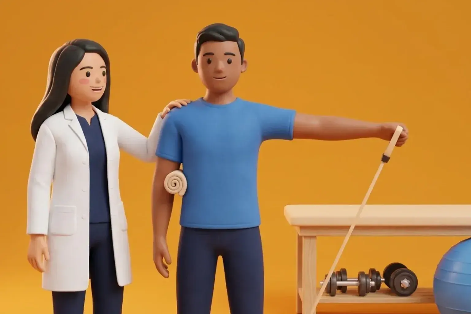
Wrapping a soft towel loosely around your elbow and securing it with tape prevents you from curling your arm tightly while sleeping. Keeping your elbow at a gentle 30 to 45 degree angle opens the nerve tunnel, restoring blood flow and stopping morning finger numbness.
Related condition guides



Related recovery guides & articles
BookPhysio.in Editorial Team
The BookPhysio.in editorial team writes and maintains this content, checking it against reliable clinical sources. Content follows our medical review policy: general information only, and no page claims a clinical review until a named registered physiotherapist has signed it off.
Read our medical review policy