
Gastrocnemius and Popliteus Muscle Crosstalk with Meniscal Horns and Joint Stability
In short
Explore the fascinating anatomical connections and functional crosstalk between the popliteus, gastrocnemius, and posterior meniscal horns.
In this guide (4 sections)
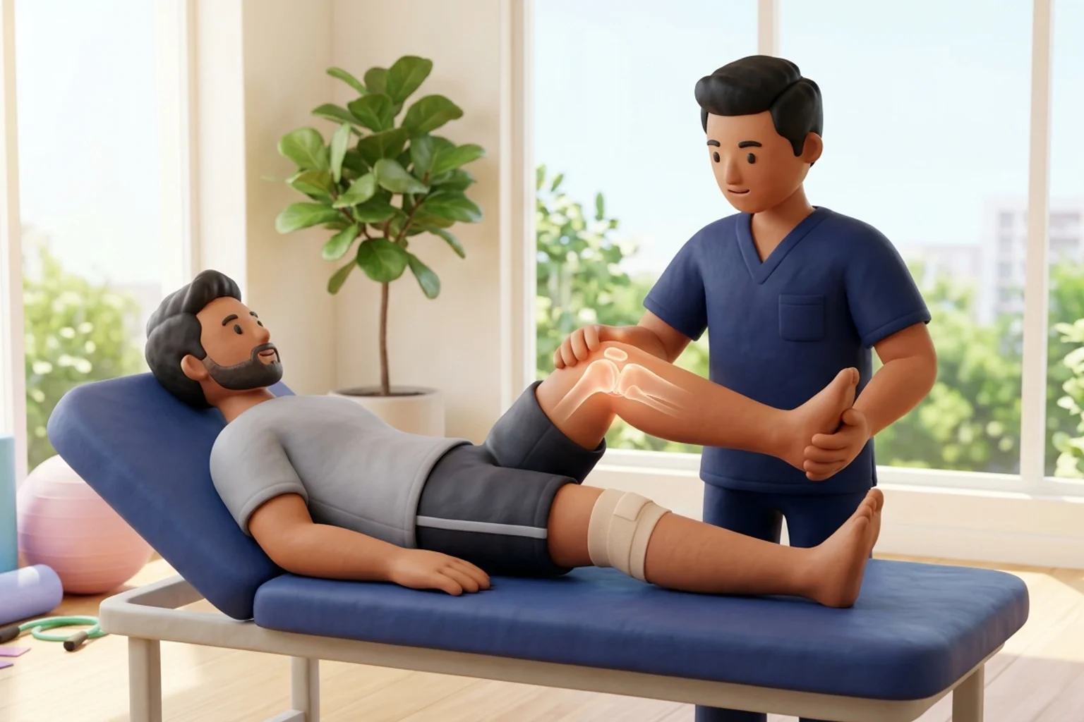
While major superficial muscle groups like the quadriceps and hamstrings receive the bulk of clinical attention, the deep musculature of the posterior knee, specifically the popliteus muscle complex and the gastrocnemius heads, exerts profound, direct anatomical control over meniscal positioning and joint mechanics. Far from being passive structures that merely respond to skeletal motion, the menisci are actively guided and retracted during joint flexion and rotation through specialized myofascial and ligamentous expansions. The popliteus muscle, through its distinct popliteomeniscal fascicles (anteroinferior, posterosuperior, and posteroinferior), directly tethers the posterior horn of the lateral meniscus. Simultaneously, the medial head of the gastrocnemius maintains intimate myofascial connections with the posterior capsule and the medial meniscal rim. Understanding this functional muscular crosstalk is essential for unravelling deep posterior knee pain and optimizing rehabilitation.
Indian Household Chores: Low Stool Scrubbing and Deep Squat Shifts
In Indian daily life, deep posterior knee muscles undergo rigorous functional loading. Performing domestic chores while seated on a low stool (patla) or shifting weight laterally while scouring cooking utensils demands fine-tuned rotational stability from the popliteus. When an individual rises from a floor mat, the medial gastrocnemius contracts forcefully to initiate plantarflexion while the knee is still partially flexed. If a patient has sustained a lateral meniscal tear involving the popliteal hiatus, or has chronic tightness in the medial calf following years of wearing flat domestic footwear, this deep muscular crosstalk becomes dysfunctional. Patients frequently complain of a deep, unreachable ache behind the knee crease that worsens during rotational domestic movements. Targeted manual therapy and specific activation drills restore harmonious muscular gliding.
Popliteomeniscal Fascicles, Hiatus Mechanics, and Lateral Meniscal Retraction
The biomechanical relationship between the popliteus tendon and the lateral meniscus is one of the most sophisticated anatomical arrangements in the musculoskeletal system. As the popliteus tendon courses obliquely through the popliteal hiatus of the joint capsule to insert into the lateral femoral condyle, it sends robust fibrous expansions, the popliteomeniscal fascicles, into the posterior horn of the lateral meniscus. When the knee initiates active flexion, the popliteus contracts, un-screwing the knee by rotating the femur externally on the fixed tibia and actively pulling the posterior horn of the lateral meniscus posteriorly and laterally. This active retraction prevents the lateral meniscus from becoming trapped and crushed between the rolling lateral femoral condyle and the tibial plateau. Disruption of these fascicles, often seen in athletic twisting injuries, causes hypermobility of the lateral meniscus, mechanical clicking, and tear propagation.
Deep Posterior Knee Rehabilitation: Popliteus Activation and Calf Eccentrics
Rehabilitating the deep posterior musculature requires exercises that integrate rotational control with controlled sagittal plane loading. To recruit the popliteus effectively, physiotherapists utilize seated internal tibial rotation drills against resistance bands, performed with the knee flexed to 90 degrees. This specific angle maximizes the mechanical advantage of the popliteus while minimizing gastrocnemius substitution. For the gastrocnemius complex, rehabilitation focuses on eccentric calf lowering over a step with both straight-knee (gastrocnemius bias) and bent-knee (soleus bias) variations. Restoring full calf flexibility eliminates equinus restrictions that would otherwise force early knee hyperextension during terminal stance, protecting the posterior capsular attachments of the menisci from repetitive tensile traction.
Seated Resisted Internal Tibial Rotation with Elastic Band
Objective: Isolate and strengthen the popliteus muscle to optimize active retraction and stability of the lateral meniscal posterior horn
- 1Sit upright on a dining chair with your affected knee bent to 90 degrees and your heel resting firmly on a smooth floor.
- 2Loop a light resistance band around the forefoot of your affected leg, anchoring the opposite end to a heavy table leg to the outside of your foot.
- 3Keeping your heel firmly anchored as a pivot point on the floor, rotate your forefoot inward toward the midline against the band resistance.
- 4Feel the deep muscle contraction behind and beneath your knee crease as your tibia rotates internally.
- 5Hold the internal rotation for 2 seconds, then slowly return to the neutral starting position over 3 seconds under elastic control.
| Muscular Structure | Meniscal Attachment / Proximity | Active Kinematic Role | Dysfunction Manifestation | Targeted Rehabilitation Drill |
|---|---|---|---|---|
| Popliteus Muscle Complex | Direct tethering via popliteomeniscal fascicles | Actively retracts lateral meniscus posterior horn in flexion | Lateral meniscus hypermobility, pseudolocking, hiatal tear | Seated resisted internal tibial rotation |
| Medial Gastrocnemius Head | Intimate fusion with posteromedial joint capsule | Dynamic posterior knee stabilization and plantarflexion drive | Posterior capsule tightness, early hyperextension shear | Eccentric calf lowering with straight knee on step |
| Plantaris Muscle | Courses adjacent to lateral head of gastrocnemius | Proprioceptive feedback and minor plantarflexion assist | Myofascial posterior knee tightness mimicking meniscal pain | Gentle posterior knee soft-tissue release |
Chronic meniscal pathology and secondary synovial effusion frequently lead to fluid accumulation in the gastrocnemio-semimembranosus bursa, forming a popliteal (Baker) cyst. If the cyst ruptures under high intra-articular pressure, synovial fluid tracks down into the calf, causing sudden severe calf pain, extensive swelling, and erythema that closely mimics deep vein thrombosis (pseudothrombophlebitis). Urgent clinical evaluation and Doppler ultrasound are required to rule out true vascular occlusion.
Frequently Asked Questions
Why does the back of my knee hurt when my meniscal tear is located inside the joint?
The menisci are connected directly to the deep muscles at the back of your knee, including the popliteus and gastrocnemius muscles. When a meniscal tear causes inflammation or mechanical friction, these deep muscles contract protectively to stabilize the joint, leading to chronic muscle tension and a deep, aching sensation in the crease behind your knee.
Do I need a doctor referral to treat a meniscal tear on BookPhysio.in?
BookPhysio.in does not require a doctor referral. Under the National Commission for Allied and Healthcare Professions (NCAHP) Act 2021, patients across India enjoy direct access to autonomous registered physiotherapists. Through BookPhysio.in, you connect directly with verified clinicians and pay per session with transparent fees, completely free from advance package commitments, commission markups, or platform subscription charges.
Related condition guides



Related recovery guides & articles
BookPhysio.in Editorial Team
The BookPhysio.in editorial team writes and maintains this content, checking it against reliable clinical sources. Content follows our medical review policy: general information only, and no page claims a clinical review until a named registered physiotherapist has signed it off.
Read our medical review policy