
Identifying Acute Plantar Fascia Rupture: The Sudden Pop, Plantar Ecchymosis, and Triage Protocols
In short
Learn how to recognize an acute plantar fascia rupture, identify the classic sudden pop and bruising, and execute immediate immobilization protocols.
In this guide (4 sections)
While chronic plantar fasciopathy is an insidious degenerative enthesopathy that develops over months, clinicians and active individuals must remain acutely vigilant for acute structural failure: complete or partial rupture of the plantar aponeurosis. An acute plantar fascial rupture is a traumatic, dramatic event characterized by an unmistakable clinical presentation. Patients almost universally report hearing or feeling a sudden, loud, sickening 'pop' or snapping sensation under the arch of the foot during a forceful push-off movement, such as sprinting, jumping, lunging in sports, or stepping off a curb. This sudden audible snap is accompanied by instantaneous, excruciating sharp pain, immediate inability to bear weight on the foot, and the subsequent appearance of extensive purple bruising (ecchymosis) spreading across the medial longitudinal arch. Distinguishing an acute rupture from routine fasciopathy is critical: attempting to walk through a rupture or performing aggressive stretching can cause retraction of the fascial stumps, permanent arch collapse, and severe long-term disability.
Barefoot Marble Floor Living, Prolonged Kitchen Standing, and Thin Flat Slippers
In Indian populations, acute plantar fascial ruptures occur through two primary clinical presentations: athletic push-off injuries and iatrogenic failures following corticosteroid injections. In urban settings, recreationally active adults playing badminton or cricket on hard courts frequently sustain acute ruptures during sudden lunges. However, an equally common scenario involves patients with pre-existing heel pain who received one or more corticosteroid injections at local clinics. Corticosteroids weaken collagen fibers and induce focal necrosis. These patients return home, feel temporary pain relief, and walk barefoot across hard marble or granite floors. A minor slip on a wet tile or a sudden step down off a stool triggers a catastrophic tear of the chemically weakened fascia. Recognizing the signs of rupture allows immediate protective action before structural arch collapse ensues.
Windlass Mechanism Strain, Calcaneal Enthesopathy, and Plantar Thickness
The pathomechanics of acute fascial rupture involve tensile forces exceeding the ultimate tensile strength of the collagen fibers. Ruptures typically occur either at the calcaneal enthesis or within the midsubstance of the central band. In patients with underlying chronic fasciopathy or prior corticosteroid exposure, the degenerative tissue possesses diminished elastic compliance, making it prone to sudden brittle failure under high-velocity loading. High-resolution diagnostic ultrasound and MRI are definitive: ultrasound reveals a complete disruption of the fibrillar architecture with an intervening anechoic fluid hematoma and retracted stump margins, while MRI demonstrates extensive hyperintense T2-weighted signal edema throughout the plantar soft tissues. A palpable focal gap or notch is frequently detectable along the medial cord during the acute phase.
Rathleff High-Load Towel Training, Low-Dye Taping, and Intrinsic Foot Doming
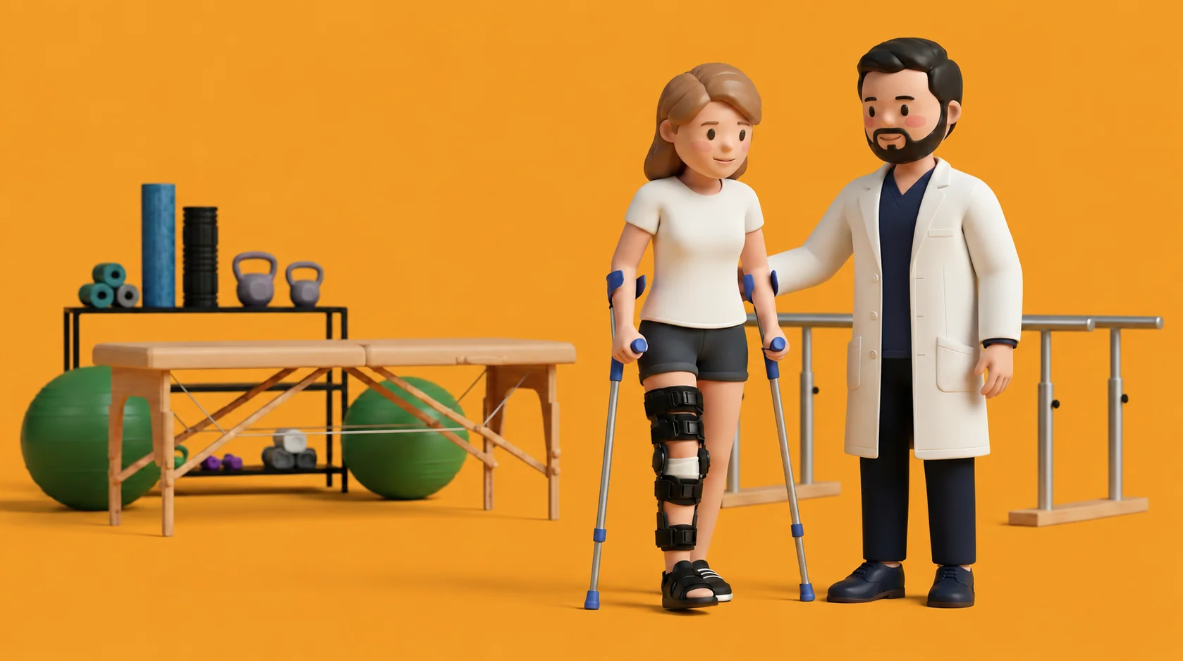
The clinical management of an acute plantar fascial rupture mandates an immediate and complete departure from chronic fasciopathy protocols. All high-load strength training (Rathleff protocol), calf stretching, and aggressive manual therapy are strictly contraindicated during the acute healing phase. Management centers on immediate non-weight-bearing immobilization in a rigid pneumatic walking boot or posterior splint with crutches for three to four weeks, allowing the disrupted collagen stumps to organize and bridge with a fibrocartilaginous scar. Once acute swelling and ecchymosis subside and clinical palpation demonstrates stable scar continuity, progressive partial weight-bearing is initiated in the boot. Rehabilitation progresses cautiously over twelve to sixteen weeks, incorporating sub-maximal isometric arch doming, gentle passive range of motion, and custom orthotic arch support to prevent permanent flatfoot deformity.
Non-Weight-Bearing Gentle Toe Wiggles and Ankle Pumps (Acute Rupture Phase)
Objective: Maintain distal circulation and prevent ankle stiffness during early cast/boot immobilization without stressing the torn fascia
- 1While resting comfortably with your injured leg elevated on pillows above heart level, remove your pneumatic boot if permitted by your clinician.
- 2Gently wiggle all five toes up and down smoothly for 30 seconds to stimulate venous return.
- 3Perform small, gentle ankle pumps up and down, keeping the movement strictly within a small, completely pain-free arc of 10 to 15 degrees.
- 4Do not allow your ankle to dorsiflex fully, and never pull your toes backward toward your shin, as this would stretch the torn fascial tissue.
- 5Carefully place your foot back into the protective pneumatic boot and secure all straps snugly.
| Clinical Parameter | Chronic Plantar Fasciopathy | Acute Plantar Fascia Partial / Complete Rupture | Immediate Clinical Triage Action |
|---|---|---|---|
| Onset & Auditory Sensation | Gradual, insidious onset over weeks; zero snapping sound or pop reported | Sudden, instantaneous tearing event; audible 'pop' or snap during push-off | Immediate non-weight-bearing; apply crutches and protective splint |
| Arch Ecchymosis & Bruising | Completely absent; skin appears normal without discoloration or swelling | Prominent purple/blue ecchymosis spreading across the medial arch within 24h | Ice, elevation, and immediate ultrasound or MRI to confirm tear extent |
| Palpation & Physical Exam | Focal point tenderness on medial tuberosity; taut continuous fascial band | Palpable soft-tissue gap or defect along the arch; exquisite diffuse tenderness | Halt all stretching and high-load exercises; refer for orthopaedic evaluation |
An acute plantar fascial rupture is an orthopaedic emergency that requires immediate immobilization. If you felt or heard a sudden pop under your arch followed by bruising and an inability to bear weight, do not attempt to walk or stretch the foot. Place the foot in a rigid walking boot or splint, use crutches to avoid all weight-bearing, and obtain immediate orthopaedic evaluation and urgent diagnostic imaging.
Frequently Asked Questions
Does an acute plantar fascia rupture require emergency surgery to sew it back together?
In the vast majority of cases, acute plantar fascia ruptures are managed non-surgically with excellent long-term recovery. Treatment involves four to six weeks of protected immobilization in a rigid walking boot, starting with crutches, allowing the torn fascial ends to bridge together with strong scar tissue. Surgery is rarely needed and is considered only if there is a severe avulsion of bone or persistent debilitating pain that fails comprehensive conservative healing.
Do I need a doctor referral to treat plantar fasciitis on BookPhysio.in?
BookPhysio.in does not require a doctor referral. Under the National Commission for Allied and Healthcare Professions (NCAHP) Act 2021, patients across India enjoy direct access to autonomous registered physiotherapists. Through BookPhysio.in, you connect directly with verified clinicians and pay per session with transparent fees, completely free from advance package commitments, commission markups, or platform subscription charges.
Related condition guides



Related recovery guides & articles
BookPhysio.in Editorial Team
The BookPhysio.in editorial team writes and maintains this content, checking it against reliable clinical sources. Content follows our medical review policy: general information only, and no page claims a clinical review until a named registered physiotherapist has signed it off.
Read our medical review policy