
Isometric Quadriceps Wall Sits with Controlled Knee Flexion Angles
In short
Master the clinical application of isometric wall sits, using controlled flexion angles to build quadriceps strength without meniscal compression.
In this guide (4 sections)
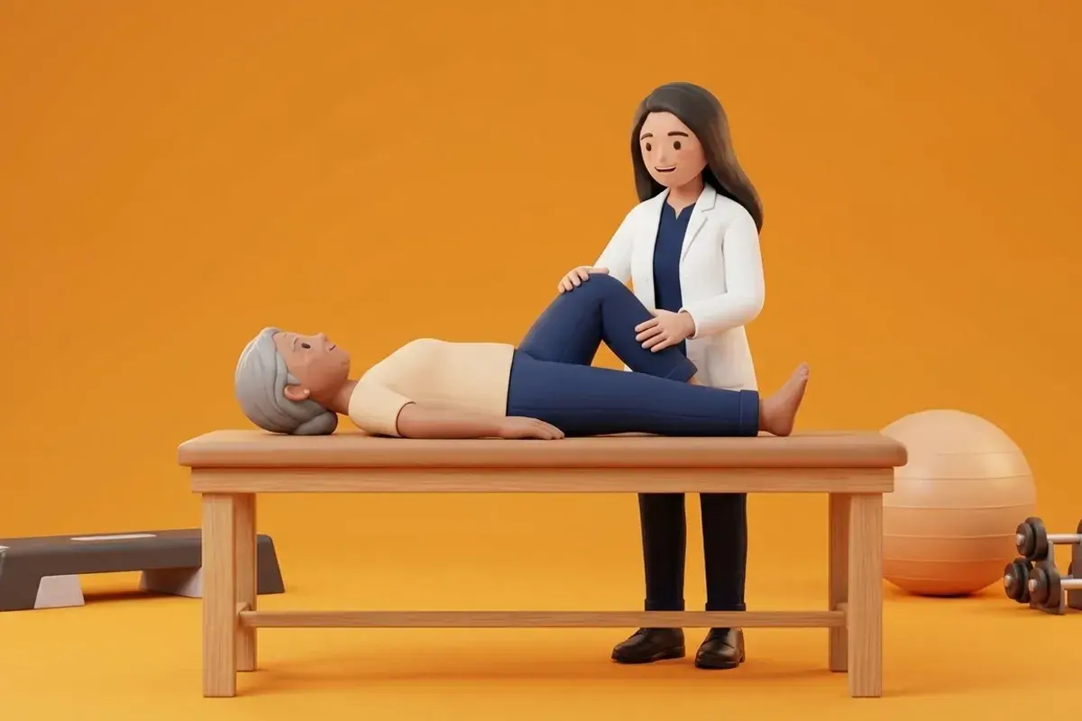
Isometric exercise, characterized by muscular force generation without changes in muscle length or joint angle, represents a foundational modality in the early and intermediate rehabilitation of meniscal injuries. Dynamic exercises, such as deep squats or lunges, involve continuous joint motion where changing contact vectors can pinch sensitive meniscal tissue between articular surfaces. In contrast, isometric quadriceps wall sits allow clinicians to select an exact, safe joint flexion angle where articular contact areas are maximized and meniscal shear is minimized. Furthermore, sports science research led by Rio and colleagues demonstrates that sustained, heavy isometric contractions exert a potent neurophysiological analgesic effect, inducing cortical motor inhibition and providing immediate pain relief that lasts for several hours following exercise.
Indian Household Simplicity: Flat Living Room Walls and Safe Kitchen Stances
In Indian domestic environments, access to specialized gym machinery or heavy weights is often limited, especially for older adults or busy homemakers. The isometric wall sit requires zero specialized equipment; any smooth, unobstructed interior wall, hallway door, or sturdy cupboard surface can serve as an effective exercise station. This makes the exercise highly practical to incorporate into everyday household routines, such as performing a set while waiting for milk to boil on the stove or during commercial breaks while watching television. Because the back is supported firmly against the wall, the risk of losing balance or falling on hard domestic tiles is virtually eliminated, providing vulnerable patients with high exercise confidence.
Angle-Dependent Tibiofemoral Physics and Shear Elimination
The clinical safety and effectiveness of the isometric wall sit depend entirely on the chosen knee flexion angle. At deep squat angles exceeding 90 degrees, tibiofemoral compressive stress soars to 5 to 7 times body weight, and the femoral condyles slide backward to compress the posterior meniscal horns. However, when a wall sit is calibrated to a shallow or moderate angle, specifically between 30 and 45 degrees of flexion, the mechanical environment is remarkably favorable. In this range, the ground reaction force vector is directed upward through the tibia, generating pure axial compression that is absorbed by the broad central body of the menisci, while anteroposterior shear forces are almost completely absent. This allows the quadriceps to generate high muscular tension (stimulating motor unit recruitment and tendon remodeling) while protecting healing tear sites.
Staged Wall Sit Prescription: Angle Progression and Bilateral Loading
Clinical execution of the wall sit requires strict adherence to angle boundaries. Patients stand with their back against a flat wall, feet positioned approximately 45 to 60 centimetres forward, hip-width apart. They slide down the wall until their knees reach the prescribed angle: 30 degrees during the acute or post-repair phase, progressing to 45 degrees, and eventually 60 degrees as tissue consolidation occurs. Holding angles beyond 60 degrees is avoided in meniscal pathology. Holds are held for 30 to 45 seconds at approximately 60 to 70 percent of maximum voluntary contraction, repeated for 4 to 5 sets with 60-second rest intervals. To enhance vastus medialis oblique (VMO) recruitment and prevent dynamic knee valgus, a soft foam ball or yoga block is placed between the knees, requiring gentle adductor co-contraction.
Supported Wall Sit at Regulated 45-Degree Flexion with Ball Squeeze
Objective: Stimulate quadriceps motor unit recruitment and analgesic cortical inhibition while maintaining zero posterior meniscal shear
- 1Stand with your back flat against a smooth, sturdy wall with your feet shoulder-width apart, approximately 50 centimetres from the baseboard.
- 2Place a soft 15-centimetre Pilates ball or folded cushion between your knees, holding it in place with gentle adductor pressure.
- 3Keeping your back and shoulders flat against the wall, slowly slide downward until your knees bend to approximately 45 degrees.
- 4Ensure your knees stay directly aligned over your second toes and do not travel forward past your shoelaces.
- 5Hold this static position firmly for 30 to 45 seconds while breathing smoothly and deeply, then press through your heels to slide back up.
| Wall Sit Flexion Angle | Meniscal Contact Region | Quadriceps Activation Level | Posterior Horn Shear Risk | Clinical Target Population |
|---|---|---|---|---|
| 30 Degrees (Shallow) | Anterior to mid-body of menisci | Moderate; excellent introductory activation | Virtually zero shear; highly safe | Acute tears, post-op repair Weeks 4 to 6 |
| 45 Degrees (Moderate) | Central broad meniscal body | High; optimal zone for muscle hypertrophy | Minimal; safe for stable fibrocartilage | Subacute tears, post-meniscectomy, conservative care |
| 60 Degrees (Advanced) | Transition toward posterior horn margin | Very high motor unit recruitment | Low to moderate; requires good tissue stability | Late-stage athletic conditioning and return to sport |
| 90 Degrees (Right Angle) | Direct posterior horn wedge zone | Maximal patellofemoral and quad load | High shear; potential tear aggravation | Contraindicated in meniscal tears and root injuries |
If a patient experiences sharp, stabbing anterior pain behind the kneecap (patellofemoral joint) rather than a muscular burning sensation in the mid-thigh during a wall sit, the knee angle is too deep or the feet are placed too close to the wall. Sliding down too far increases patellofemoral joint reaction forces exponentially. Clinicians should immediately reduce the angle by 10 to 15 degrees and ensure the shins remain strictly vertical.
Frequently Asked Questions
Why does my knee feel less painful after holding a wall sit for 45 seconds?
Sustained isometric muscle contractions trigger a proven neurological pain-relieving phenomenon called exercise-induced hypoalgesia. Contracting your quadriceps firmly without moving the joint stimulates sensory mechanoreceptors in your tendons, which sends inhibitory signals to your spinal cord and brain, dampening pain signals for several hours.
Do I need a doctor referral to treat a meniscal tear on BookPhysio.in?
BookPhysio.in does not require a doctor referral. Under the National Commission for Allied and Healthcare Professions (NCAHP) Act 2021, patients across India enjoy direct access to autonomous registered physiotherapists. Through BookPhysio.in, you connect directly with verified clinicians and pay per session with transparent fees, completely free from advance package commitments, commission markups, or platform subscription charges.
Related condition guides



Related recovery guides & articles
BookPhysio.in Editorial Team
The BookPhysio.in editorial team writes and maintains this content, checking it against reliable clinical sources. Content follows our medical review policy: general information only, and no page claims a clinical review until a named registered physiotherapist has signed it off.
Read our medical review policy