
Kabaddi Knee Multi-Ligament Tear: ACL, MCL and PLC Tackle Rehabilitation
In short
Violent defensive chain tackles and ankle catches in kabaddi exert massive rotational valgus shear, causing catastrophic multi-ligament knee disruptions.
In this guide (7 sections)
Kabaddi is an elite collision sport combining elements of wrestling, sprint agility, and martial combat. During a raid, a raider crossing the baulk line faces coordinated defensive formations such as the chain tackle, thigh hold, and ankle catch. Multi-ligament knee injuries - characterized by simultaneous rupture of the anterior cruciate ligament (ACL), medial collateral ligament (MCL), and posterolateral corner (PLC) - represent the most severe orthopaedic trauma encountered in the sport. The injury results from high-velocity impact where the raider's planted foot is trapped while multiple defenders drive through the knee, exerting catastrophic valgus, hyperextension, and external rotation shear forces.
Domestic Indian Sports Reality and Field Conditions
In traditional rural Indian kabaddi, matches take place on soft clay or mud akhadas prepared with sand, cow dung ash, and mustard oil to provide traction while reducing skin abrasions. However, the modern sport has largely transitioned to dense synthetic EVA foam mats used in the Pro Kabaddi League and national championships. While synthetic mats offer consistent traction, they lack rotational give. When an ankle catch locks a raider's foot against the rubberized mat, the knee absorbs all twisting momentum from multiple converging defenders weighing over 80 kg each. In village and district tournaments, field-side emergency stabilization is rarely available, leading to neglected ligamentous knee dislocations.
Biomechanics and Pathomechanics of Injury
The pathomechanics of a multi-ligament knee injury in kabaddi typically follow a 'dashboard' or 'unhappy triad' variation under multi-directional loading. When a defender tackles the raider laterally while the foot is fixed in external rotation, the superficial and deep MCL fibers yield first under extreme valgus tension. As valgus displacement exceeds 15 degrees, the ACL tears as it impinges against the intercondylar notch. Continued hyperextension and tibial external rotation ruptures the structures of the PLC, including the fibular collateral ligament (FCL), popliteus tendon, and popliteofibular ligament. This triad results in severe multi-planar rotational instability and carries a high risk of common peroneal nerve stretch injury.
Clinical Assessment and Provocative Special Tests
- Lachman and Valgus Stress Tests at 0° and 30°: Evaluates ACL and MCL integrity. Gross medial joint space opening (>10 mm) at 0 degrees extension confirms combined MCL and posterior oblique/cruciate disruption.
- The Dial Test at 30° and 90° Knee Flexion: The patient lies prone while the examiner passively externally rotates both tibias. Increased external rotation (>10 degrees compared to uninjured side) at 30 degrees indicates isolated PLC injury; increased rotation at both 30 and 90 degrees confirms combined PLC and PCL tears.
- Neurovascular Assessment (Peroneal Nerve and Distal Pulses): Assessing active ankle dorsiflexion, sensation in the first web space, and palpating the dorsalis pedis and posterior tibial pulses is mandatory to rule out common peroneal neuropraxia and popliteal artery intimal disruption.
Stage-by-Stage Functional Return Protocol
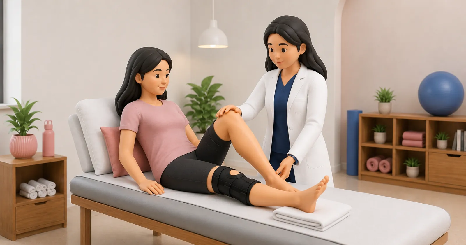
- Phase 1: Postoperative Protection and Graft Healing (Weeks 0 to 6): Maintain hinged knee brace locked in extension for initial 2 to 4 weeks, with non-weight-bearing crutch ambulation. Initiate passive knee flexion limited to 90 degrees. Perform quad sets, patellar mobilizations, and ankle pumps to prevent arthrofibrosis.
- Phase 2: Progressive Loading and Closed-Chain Conditioning (Weeks 6 to 16): Unlock brace for progressive weight-bearing. Introduce closed-chain leg presses (0 to 60 degrees), stationary cycling, and seated calf raises. Emphasize quadriceps hypertrophy without inducing anterior or posterior tibial shear.
- Phase 3: Neuromuscular Re-education and Perturbation (Months 4 to 8): Advance to bilateral and single-leg squats, Romanian deadlifts, and wobble board balance drills. Incorporate multi-directional perturbation training to recondition dynamic joint position sense.
- Phase 4: Sport-Specific Agility and Contact Deceleration (Months 9 to 14): Progress through linear sprinting, zigzag agility, defensive footwork (toe touch drills, dubki evasive movements), and controlled grappling re-entry. Clearance requires Limb Symmetry Index (LSI) >90% and psychological readiness.
Prone Isometric Hamstring-Glute Co-contraction with Banded Resistance
Objective: Builds active dynamic stabilization of the posterior and lateral knee capsule without placing anterior cruciate grafts under shear.
- 1Lie face down on a treatment mat with a light resistance band looped around your ankles.
- 2Squeeze your gluteal muscles firmly to lock your pelvis against the plinth.
- 3Slowly flex the injured knee against the band resistance to approximately 45 degrees.
- 4Hold this isometric contraction statically for 10 seconds, focusing on recruiting the hamstring bellies.
- 5Lower smoothly back to the floor under control.
Multi-Ligament Classification and Reconstruction Pathways
| Classification | Ligamentous Structures Disrupted | Vascular / Nerve Risk | Return to Contact Sport Timeline | |
|---|---|---|---|---|
| KD-I | Cruciate (ACL or PCL) + One Collateral | Low (<5%) | 6 to 9 months | |
| KD-II | Bicruciate (ACL + PCL) without collateral | Moderate (10% - 15%) | 9 to 12 months | |
| KD-III | Bicruciate (ACL + PCL) + MCL or PLC | High (20% - 25% popliteal artery risk) | 12 to 16 months | |
| KD-IV | Bicruciate (ACL + PCL) + Both MCL & PLC | Very High (>40% vascular, 25% peroneal nerve) | 15 to 18 months (recreational focus) |
Red Flag Signs Requiring Immediate Medical Referral
Diminished or absent pedal pulses, a cold, pale, or cyanotic foot, ankle-brachial index (ABI) <0.9, or rapidly progressive leg swelling following knee dislocation indicates traumatic popliteal artery disruption, constituting a limb-threatening surgical emergency requiring emergency vascular revascularization within 6 to 8 hours.
Frequently Asked Questions
Can an athlete return to professional kabaddi after multi-ligament knee surgery?
While challenging, elite kabaddi athletes can return to competition following multi-ligament reconstruction if they achieve full structural graft healing, restore 90% limb strength symmetry, and complete extensive contact reconditioning over 12 to 14 months.
Why is the posterolateral corner (PLC) frequently missed in clinical exams?
Isolated ACL or MCL laxity often masks underlying PLC instability. If the examiner fails to perform the Dial test or assess external rotational recurvatum, untreated PLC deficiency will cause catastrophic overload and failure of the reconstructed ACL graft.
Related condition guides



Related recovery guides & articles
BookPhysio.in Editorial Team
The BookPhysio.in editorial team writes and maintains this content, checking it against reliable clinical sources. Content follows our medical review policy: general information only, and no page claims a clinical review until a named registered physiotherapist has signed it off.
Read our medical review policy