
L3-L4 Disc Herniation and L4 Radiculopathy: Femoral Nerve Stretch Test and Quadriceps Weakness
In short
Understand upper lumbar L3-L4 disc herniations, L4 nerve root compression, quadriceps weakness, patellar reflex loss, and reverse Lasegue testing.
In this guide (4 sections)
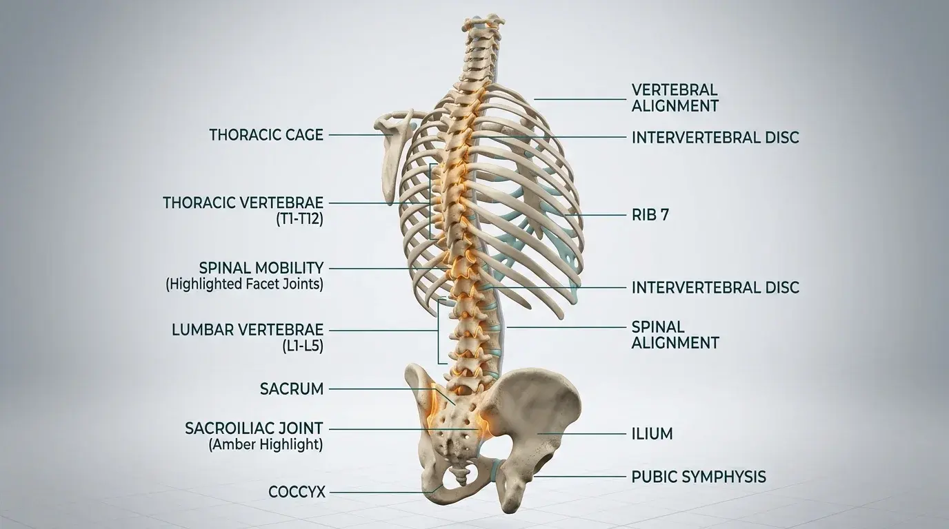
While the vast majority of lumbar disc herniations occur at the lower two spinal levels, approximately 5 percent of clinical cases arise at the upper lumbar segments, predominantly at L3-L4. Herniation at this level compromises the L4 nerve root, producing a clinical radiculopathy that differs fundamentally from classic posterior sciatica. Instead of radiating down the back of the thigh to the heel, L4 radicular pain tracks across the anterior and anteromedial aspect of the thigh, wraps around the front of the knee, and extends down the medial shin to the medial malleolus. Because the L4 root forms a critical component of the femoral nerve and lumbar plexus, its compression impairs the quadriceps femoris muscle, leading to sudden knee buckling and instability. Clinicians must recognize these distinctive features, as standard straight leg raising tests are often completely negative in upper lumbar radiculopathy.
Two-Wheeler Pothole Shocks, Indian Squatting Commode Hazards, and IT Chair Stress
In domestic environments across India, L4 radiculopathy introduces acute safety risks, particularly involving stair navigation and low seating. In multi-story residential buildings without elevators, descending stairs requires strong eccentric quadriceps control; patients with L4 weakness experience sudden knee buckling, creating high risks of traumatic falls. The common household practice of sitting on low stools, such as a peeda or wooden patla while washing utensils or cooking, becomes impossible because getting up requires explosive knee extension strength. In traditional squatting toilets, the deep flexion puts intense strain on weakened quadriceps, forcing individuals to pull themselves up using walls or doorframes. Furthermore, sitting cross-legged on the floor for extended family prayers applies sustained traction across the femoral nerve distribution, worsening anterior thigh burning.
Annular Disruption, Chemical Radiculitis, and Dermatomal Nerve Root Compression
The L4 nerve root exits through the L4-L5 neural foramen, but in a paracentral L3-L4 disc herniation, it is compressed as a traversing root within the lateral recess. Motor fibers of the L4 root innervate the quadriceps femoris, tibialis anterior, and tensor fasciae latae. Significant compression reduces motor unit recruitment to the vastus medialis and rectus femoris, compromising terminal knee extension. Concurrently, the afferent and efferent limbs of the patellar tendon reflex arc are interrupted, resulting in a diminished or absent knee jerk. Because the femoral nerve runs anteriorly through the pelvis and femoral triangle, standard straight leg raise tests fail to tension the L4 root. Instead, the prone knee bend test, also known as the Femoral Nerve Stretch Test or Reverse Lasegue sign, exerts longitudinal traction on the nerve, reproducing sharp anterior thigh pain.
McKenzie Centralization, Neural Dynamic Sliders, and Graded Lumbar Mobilization
Physiotherapy rehabilitation for L3-L4 disc lesions focuses on gentle decompression, quadriceps motor re-education, and femoral nerve mobilization. Prone positioning must be introduced cautiously, as excessive lumbar extension combined with hip extension can pinch the upper lumbar foramen. Controlled prone lying with a pillow placed under the abdomen provides an initial neutral offloading posture. Once acute pain subsides, femoral nerve sliders are performed in the side-lying position, alternating knee flexion with cervical extension to glide the nerve smoothly through the psoas muscle and femoral canal without excessive stretch. Closed-chain quadriceps strengthening begins with shallow, supported wall slides and step-ups to rebuild functional knee stability.
Side-Lying Femoral Nerve Slider with Coordinated Cervical Motion
Objective: Mobilize the L4 nerve root and femoral nerve through the retroperitoneal space without aggravating radicular tension
- 1Lie on your unaffected side on a supportive bed or mat, bending your bottom hip and knee to 90 degrees for pelvic stability.
- 2Keep your head supported on a comfortable pillow so your neck remains in a neutral alignment.
- 3Slowly bend your upper (affected) knee toward your gluteal muscles while simultaneously extending your neck backward toward the ceiling.
- 4Hold this end position for 1 second, feeling a gentle motion through the front of your thigh without reproducing sharp pain.
- 5Smoothly reverse the movement by straightening your knee forward while simultaneously flexing your chin down toward your chest.
| Clinical Feature | L4 Radiculopathy (L3-L4 Disc) | L5 Radiculopathy (L4-L5 Disc) | S1 Radiculopathy (L5-S1 Disc) |
|---|---|---|---|
| Primary Pain Distribution | Anterior thigh, across the patella, into the medial lower leg and medial malleolus | Posterolateral thigh, lateral knee, anterior shin, and dorsum of foot to great toe | Direct posterior thigh, popliteal fossa, posterior calf, heel, and lateral foot border |
| Primary Motor Weakness | Quadriceps femoris (knee extension) and partial tibialis anterior weakness | Extensor hallucis longus (big toe lift) and hip abductors (gluteus medius) | Gastrocnemius and soleus (plantarflexion) and peroneus longus/brevis |
| Reflex Abnormality | Diminished or absent patellar tendon reflex (knee jerk) | No standard deep tendon reflex (normal patellar and Achilles reflexes) | Diminished or absent Achilles tendon reflex (ankle jerk) |
| Primary Neurodynamic Test | Positive Prone Knee Bend (Femoral Nerve Stretch / Reverse Lasegue) | Positive Straight Leg Raise (SLR) and Slump test | Positive Straight Leg Raise (SLR) and Slump test |
Upper lumbar disc herniations occupy an area of the spinal canal where the thecal sac contains numerous descending nerve roots. Rapid onset of bilateral quadriceps paralysis, sudden inability to stand upright, or loss of urinary control necessitates emergency neurosurgical evaluation within hours.
Frequently Asked Questions
Why does my knee keep giving way when my doctor said the problem is in my back?
Your L3-L4 disc herniation pinches the L4 nerve root, which powers the quadriceps muscles on the front of your thigh. The quadriceps act as the primary stabilizer to keep your knee locked while walking or descending stairs. When the nerve is compressed, muscle activation drops, causing your knee to buckle unexpectedly.
Do I need a doctor referral to treat a herniated disc on BookPhysio.in?
BookPhysio.in does not require a doctor referral. Under the National Commission for Allied and Healthcare Professions (NCAHP) Act 2021, patients across India enjoy direct access to autonomous registered physiotherapists. Through BookPhysio.in, you connect directly with verified clinicians and pay per session with transparent fees, completely free from advance package commitments, commission markups, or platform subscription charges.
Related condition guides



Related recovery guides & articles
BookPhysio.in Editorial Team
The BookPhysio.in editorial team writes and maintains this content, checking it against reliable clinical sources. Content follows our medical review policy: general information only, and no page claims a clinical review until a named registered physiotherapist has signed it off.
Read our medical review policy