
Lumbar Spinal Fusion (TLIF / PLIF): Bone Graft Incorporation and Neutral Spine Rehabilitation
In short
Lumbar spinal fusion creates permanent rigid bone union between vertebrae. Learn bone graft incorporation phases, hardware protection, and neutral spine core stabilization.
In this guide (5 sections)
The Biology and Biomechanics of Lumbar Spinal Fusion
Lumbar spinal fusion is indicated for mechanical instability, high-grade spondylolisthesis, recurrent disc herniations, and degenerative scoliosis.
Procedures such as Transforaminal Lumbar Interbody Fusion (TLIF) and Posterior Lumbar Interbody Fusion (PLIF) excise the degenerative disc, insert an interbody spacer cage packed with bone graft, and secure the motion segment using bilateral titanium pedicle screws and connecting rods.
It is essential to understand that pedicle screws and rods are merely temporary internal fixation splints; permanent stability requires biological bone union (arthrodesis) between the vertebral endplates, a process requiring 3 to 12 months of osteoblastic remodeling.
Bone Graft Incorporation Phases (Months 1 to 12)
Physiotherapy progression must closely mirror biological bone healing to prevent hardware loosening, screw pull-out, or pseudarthrosis (non-union).
| Post-Operative Timeline | Bone Graft Status & Union | Biomechanical Vulnerability | Physiotherapy Rehabilitation Focus | |
|---|---|---|---|---|
| Phase 1 (Weeks 0 to 6) | Inflammatory cellular recruitment; clot organization | Hardware carries 100% of mechanical load; vulnerable to shear | Log-rolling, walking intervals, strict BLT precautions, LSO brace. | |
| Phase 2 (Weeks 7 to 12) | Early osteoid matrix deposition and early callus bridging | Intermediate stiffness; early interbody bone bridging | Wean brace; isometric core in neutral; stationary cycling; step-ups. | |
| Phase 3 (Months 3 to 6) | Woven bone transforms into lamellar trabecular bone | Solid trabecular bone bridging visible on CT / radiographs | Hip mobility, hamstring flexibility, light resistance training. | |
| Phase 4 (Months 6 to 12) | Complete solid mature cortical and trabecular arthrodesis | Fused segment is fully rigid; load transferred to bone | Full functional conditioning; avoid extreme end-range lumbar hyperextension. |
The Neutral Spine Core Stabilization Hierarchy
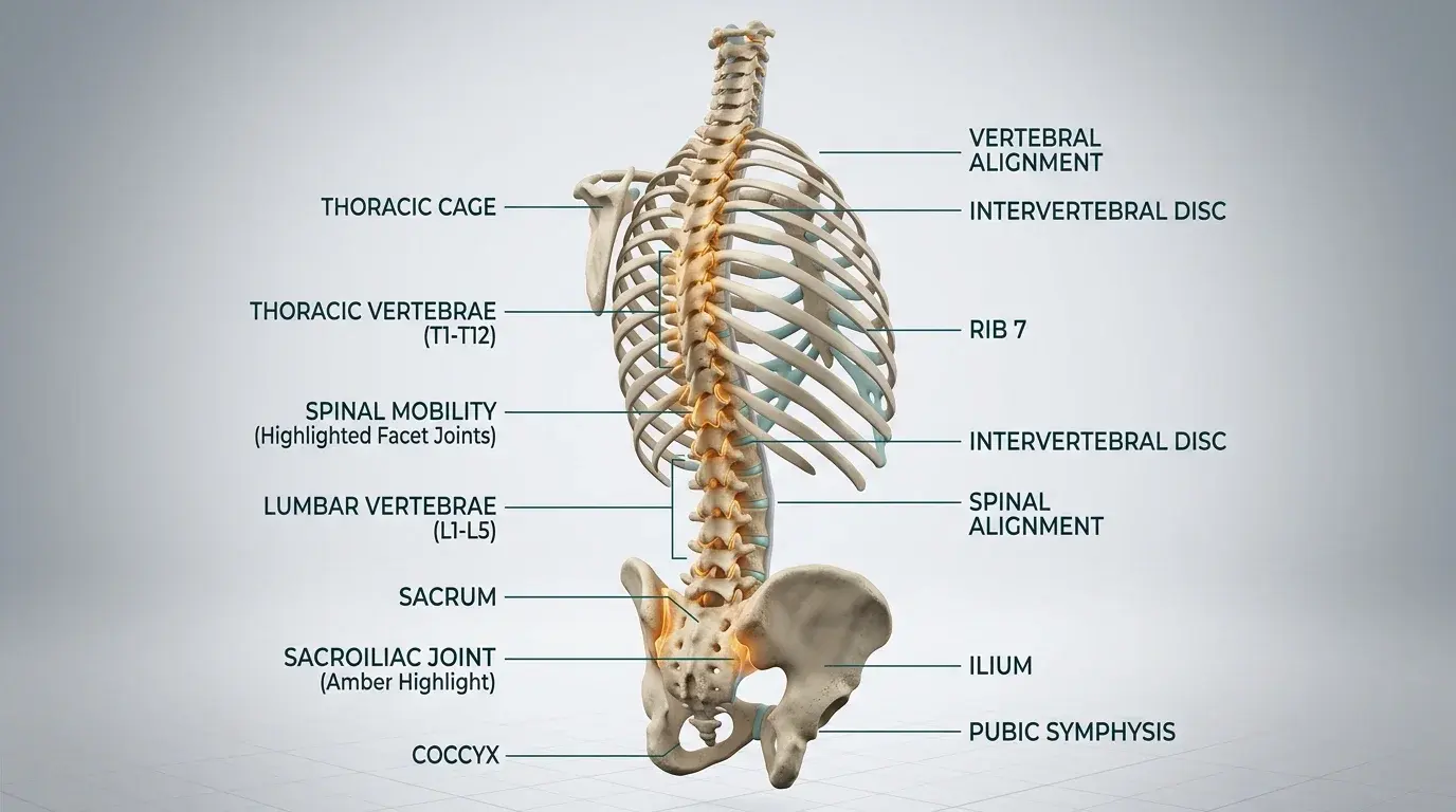
Because the fused vertebral segment can no longer move, adjacent mobile segments (particularly the level immediately above the fusion) must absorb increased rotational and flexion stresses, predisposing to Adjacent Segment Disease (ASD).
Rehabilitation trains the patient to maintain a strict 'neutral spine' during all functional movements, utilizing hip mobility rather than spinal bending.
- The Hip Hinge Re-education: Train bending strictly through the acetabulofemoral hip joints with a flat back, maintaining a wooden dowel contact along the sacrum, thoracic spine, and back of head.
- The McGill Big Three (Spine-Sparing Core): Perform modified curl-ups with hands under lumbar spine, side-planks on knees, and bird-dog quadruped reaches to build core endurance without spinal motion.
- Lumbosacral Orthosis (LSO) Brace Protocol: Wear a semi-rigid LSO brace during all out-of-bed ambulation and transfers for the first 6 to 8 weeks, removing it when lying supine.
- Hip Flexor and Hamstring Mobility: Gently stretch tight iliopsoas and hamstrings to eliminate pelvic tilting that strains the adjacent lumbar motion segments.
Indian Household Realities: Floor Chores and Commute
In India, domestic tasks (sweeping with a jhadu, washing clothes on low boards) and bumpy two-wheeler auto-rickshaw commutes exert high vibrational and rotational shear on healing fusion cages.
Physiotherapists mandate long-handled cleaning tools, elevated chairs, and avoiding two-wheeler travel for a minimum of 3 to 6 months post-fusion.
Frequently Asked Questions
What is Adjacent Segment Disease (ASD)?
Fusing a motion segment increases mechanical stiffness, causing the mobile vertebral disc immediately above or below the fusion to absorb disproportionately high compensatory flexion and rotational stresses, predisposing to accelerated degeneration over 5 to 10 years.
How do you know if a spinal fusion has solidly fused?
Solid bony union is confirmed on follow-up X-rays or thin-cut CT scans at 6 to 12 months, showing continuous trabecular bone bridging through and around the interbody cage without radiolucent halos around pedicle screws.
Can I ever bend down to touch my toes after a spinal fusion?
Patients with a 1-level fusion (e.g., L4-L5) can easily reach their toes by hinging at the hips. However, deliberately flexing the lumbar spine to end-range is discouraged; patients are taught lifelong hip-hinging mechanics to protect adjacent discs.
Related condition guides

Related recovery guides & articles
BookPhysio.in Editorial Team
The BookPhysio.in editorial team writes and maintains this content, checking it against reliable clinical sources. Content follows our medical review policy: general information only, and no page claims a clinical review until a named registered physiotherapist has signed it off.
Read our medical review policy