
Manual Therapy Posterior Talar Glides: Restoring Ankle Dorsiflexion Range Post-Fracture
In short
Master manual therapy posterior talar glides, Maitland oscillations, and Mulligan mobilization with movement to restore full dorsiflexion after ankle fracture.
In this guide (4 sections)
Restoration of terminal ankle dorsiflexion represents the primary determinant of successful functional rehabilitation following surgical fixation or conservative management of ankle fractures. Patients lacking adequate dorsiflexion exhibit severe biomechanical compensations, including early heel-off, knee hyperextension, contralateral pelvic drop, and pronatory collapse of the midfoot during the stance phase of walking. In the post-fracture ankle, simple stretching of the gastrocnemius-soleus complex frequently fails because the primary mechanical restriction is intra-articular rather than purely muscular. Because the convex talar dome must translate posteriorly within the concave mortise during dorsiflexion, capsular adhesions and anterior scar tissue physically block this movement. Manual therapy employing posterior talar glides directly restores this essential arthrokinematic slide, unlocking physiological range of motion.
Confronting Indian Squatting Commode Strains and Kitchen Kneeling Demands
In Indian households, the functional necessity for deep dorsiflexion is pervasive. Utilizing traditional Indian squatting toilets requires an extraordinary thirty-five to forty degrees of closed-chain ankle dorsiflexion as the center of gravity drops deeply over the feet; a patient with stiff posterior talar translation cannot perform this movement without tipping backward or placing intolerable strain on healing malleolar screws. Similarly, domestic tasks such as grinding spices on floor-mounted stones, rolling flatbreads on low stools, or performing morning yoga postures (asanas) demand deep ankle flexion. Without adequate posterior talar glides, attempting these domestic activities causes severe anterior ankle joint pinching, compensatory external rotation of the foot, and abnormal stress across healing surgical hardware.
Arthrokinematic Mechanics, Maitland Grades, and Mulligan Dynamic Belting
The convex-on-concave kinematics of the talocrural joint dictate that anterior rolling of the talus must be accompanied by a simultaneous posterior glide to maintain joint congruency within the mortise. When capsular fibrosis impedes this posterior translation, the talus impinges against the anterior tibial plafond. Manual therapy directly counteracts this pathology through two primary techniques: Maitland passive oscillatory mobilizations and Mulligan Mobilization with Movement (MWM). Maitland Grade III (large-amplitude oscillations up to the tissue resistance limit) and Grade IV (small-amplitude oscillations at the end-range) mechanically elongate contracted posterior capsular fibers and stimulate articular mechanoreceptors to inhibit nociceptive guarding. Mulligan MWM combines this manual glide with active patient movement, using a mobilization strap to sustain a posterior talar force while the patient actively lunges forward, retraining the neuromuscular system to execute proper arthrokinematic tracking under functional loading.
Clinical Application Protocols, Patient Positioning, and Progressive Loading
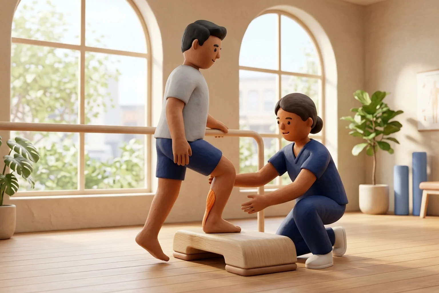
Manual posterior talar glides are implemented through structured progression. Initial treatments place the patient in a supine position with the knee flexed to relax the gastrocnemius. The clinician stabilizes the posterior distal tibia and fibula against the plinth with one hand while the web space of the treating hand contacts the anterior neck of the talus, applying a localized posterior and slightly inferior force. Once passive joint play improves, therapy transitions to weight-bearing Mulligan MWM. The patient places their foot on a treatment stool with a padded mobilization belt looped over the anterior talar neck and around the clinician's pelvis. As the clinician leans back to apply a sustained posterior glide, the patient lunges the knee forward over the toes. This is repeated for three sets of ten repetitions, immediately followed by functional stair-climbing or squatting exercises to lock in the mobility gains.
Self-Mobilization Posterior Talar Glide with Elastic Band in Half-Kneeling
Objective: Utilize elastic resistance to sustain a posterior talar glide during active functional weight-bearing dorsiflexion
- 1Adopt a half-kneeling position on a carpeted floor, placing your injured foot forward with knee bent at ninety degrees and your rear knee resting on a comfortable cushion.
- 2Secure one end of a heavy-resistance elastic mobilization band to a heavy, immovable anchor (such as a door frame or sofa leg) directly behind you at floor level.
- 3Loop the open loop of the band around your front ankle, ensuring the band rests directly below the malleoli across the anterior neck of the talus, not up on the shin.
- 4Step forward until you feel a firm, strong backward pull from the band drawing your talus directly backward toward the wall behind you.
- 5Keeping your front heel firmly glued to the floor, slowly lunge your knee forward directly over your toes into maximum comfortable dorsiflexion, hold for five seconds, then return.
| Technique Framework | Patient Position & Setup | Force Vector & Application | Clinical Indication & Timing |
|---|---|---|---|
| Maitland Grade III Glide | Supine with knee flexed 45 deg, heel resting off table edge | Large-amplitude oscillatory glide into posterior capsular resistance | Initiated at week 6 to 8 post-ORIF to treat capsular stiffness |
| Maitland Grade IV Glide | Supine, neutral ankle, manual grip over anterior talus neck | Small-amplitude oscillatory glide at absolute end of available range | Applied at weeks 8 to 12 to eliminate stubborn terminal restriction |
| Mulligan MWM with Strap | Half-kneeling or standing with foot on stool; belt over anterior talus | Sustained posterior glide maintained during active forward lunge | Initiated once full weight-bearing is cleared to restore functional gait |
When executing posterior talar glides, exact hand or belt placement over the neck of the talus is essential. Placing the contact point too high against the anterior distal tibia simply compresses the tibiofibular joint without gliding the talus, while placing it too low across the midfoot compresses the navicular bone. Never apply direct, heavy manual pressure over recent surgical incision lines or prominent screw heads.
Frequently Asked Questions
Can I do posterior talar glides at home by myself, or do I need a physiotherapist?
Your physiotherapist will initially perform manual glides in the clinic to accurately mobilize the specific stiff planes of your joint capsule without stressing your surgical hardware. Once your joint play has improved and you have learned proper band placement below the ankle bones, your physiotherapist will prescribe safe self-mobilization drills using an elastic resistance band at home.
Do I need a doctor referral to rehabilitate an ankle fracture on BookPhysio.in?
BookPhysio.in does not require a doctor referral. Under the National Commission for Allied and Healthcare Professions (NCAHP) Act 2021, patients across India enjoy direct access to autonomous registered physiotherapists. Through BookPhysio.in, you connect directly with verified clinicians and pay per session with transparent fees, completely free from advance package commitments, commission markups, or platform subscription charges.
Related condition guides



Related recovery guides & articles
BookPhysio.in Editorial Team
The BookPhysio.in editorial team writes and maintains this content, checking it against reliable clinical sources. Content follows our medical review policy: general information only, and no page claims a clinical review until a named registered physiotherapist has signed it off.
Read our medical review policy