
Microdiscectomy Post-Operative Physiotherapy Protocol: Weeks One to Twelve Recovery Phasing
In short
Master the three-phase post-operative rehabilitation protocol following lumbar microdiscectomy, balancing neural mobility, scar remodeling, and recurrence prevention.
In this guide (4 sections)
Lumbar microdiscectomy remains the gold standard surgical procedure for patients with persistent, debilitating radiculopathy or progressive motor deficits unresponsive to conservative care. Utilizing an operating microscope or high-resolution loupe magnification through a small skin incision (typically 2 to 3 centimeters), the spine surgeon removes only the ruptured disc fragment that is compressing the nerve root, preserving the majority of the healthy disc and facet joint anatomy. However, successful surgical decompression represents only the structural beginning of recovery. A structured, phased post-operative physiotherapy protocol is essential to remodel surgical scar tissue, prevent epidural fibrosis, restore neural mobility, and re-educate the deep spinal stabilizing musculature. A systematic twelve-week rehabilitation pathway safely bridges the transition from initial wound healing to full functional restoration.
Two-Wheeler Pothole Shocks, Indian Squatting Commode Hazards, and IT Chair Stress
In the post-operative period within Indian homes, patients face unique environmental challenges that can threaten the surgical site if not properly managed. During the initial six weeks, the surgical annulotomy defect remains vulnerable to re-herniation under sudden flexion or twisting loads. The traditional habit of sitting on low floor rugs for meals or using Indian squatting toilets must be strictly avoided; households should arrange a raised western toilet chair or commode attachment prior to surgery. When rising from bed, patients must abandon the common habit of curling forward and instead employ the log-roll technique, keeping the shoulders and hips aligned. Commuting on motorcycles or auto-rickshaws must be strictly prohibited during the first eight weeks, as sudden vertical jolts can disrupt early collagen healing across the surgical incision.
Annular Disruption, Chemical Radiculitis, and Dermatomal Nerve Root Compression
The biological healing following microdiscectomy progresses through three distinct phases: the inflammatory phase (weeks one to two), the fibroblastic proliferation phase (weeks three to six), and the collagen remodeling phase (weeks seven to twelve and beyond). During surgery, the small annulotomy window through which the fragment was extracted is left open to heal by secondary intention, forming a vascularized fibrous plug. If excessive hydrostatic pressure is generated during the early weeks, nuclear fragments remaining inside the disc can extrude through this healing defect, resulting in recurrent disc herniation (which occurs in roughly 5 to 10 percent of cases). Concurrently, surgical trauma triggers postoperative epidural hematoma formation and fibroblast proliferation, which can tether the freshly decompressed nerve root to adjacent bony lamina if gentle neural gliding is not introduced.
McKenzie Centralization, Neural Dynamic Sliders, and Graded Lumbar Mobilization
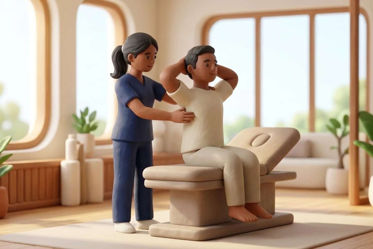
Post-microdiscectomy rehabilitation is strictly staged to respect tissue healing. In Phase 1 (weeks 1 to 2), the focus is on gentle wound healing, log-rolling education, frequent short walks (5 to 10 minutes), and gentle supine ankle pumps and sciatic nerve sliders to prevent perineural adhesion. In Phase 2 (weeks 3 to 6), formal physiotherapy commences, introducing isometric transverse abdominis activation, quadruped bird-dog drills, and hip hinge re-education, while strictly enforcing the BLT restrictions: no bending, no lifting over 5 kilograms, and no twisting. In Phase 3 (weeks 7 to 12), resistance training begins with progressive cable rows, hip thrusters, suitcase carries, and gradual introduction of controlled spinal flexion and rotation, rebuilding full physical capacity for occupational and recreational demands.
Post-Operative Quadruped Bird-Dog with Spine-Neutral Bracing
Objective: Re-educate the multifidus and transverse abdominis to stabilize the lumbar spine without applying torque to the healing annulotomy
- 1Position yourself on hands and knees on a firm, comfortable exercise mat, with wrists directly under shoulders and knees under hips.
- 2Find your neutral spine position, maintaining a slight, natural curve in your lower back without sagging or rounding.
- 3Gently draw your navel toward your spine to engage your deep core, breathing naturally throughout the exercise.
- 4Slowly reach your right arm straight forward while simultaneously extending your left leg straight back, keeping your foot flexed.
- 5Hold the extended position for 3 seconds, keeping your hips and shoulders completely level with the floor, then return smoothly and switch sides.
| Protocol Phase | Primary Rehabilitation Goals | Permitted Therapeutic Interventions | Movement Precautions and Restrictions |
|---|---|---|---|
| Phase 1: Protection (Weeks 1 to 2) | Wound healing, pain management, prevention of deep vein thrombosis, and early neural mobilization | Frequent short walking intervals (5-10 mins), log rolling, supine ankle pumps, gentle sciatic sliders | Strict BLT rule: no forward bending, no lifting over 3 kg, no spinal twisting, no sitting over 20 mins |
| Phase 2: Activation (Weeks 3 to 6) | Deep core activation, hip mobility restoration, postural re-education, and endurance walking | Quadruped bird-dog, dead bugs, supine pelvic tilts, glute bridges, stationary cycling on upright bike | Avoid loaded end-range spinal flexion; avoid sitting on low sofas; lift limit capped at 5 kg |
| Phase 3: Strengthening (Weeks 7 to 12) | Restoration of functional spinal resilience, progressive resistance training, and vocational return | Goblet squats, suitcase carries, cable rows, progressive hip hinges, graded return to driving | Maintain strict hip hinge mechanics; avoid ballistic lifting or high-impact running until week 12 |
Although microdiscectomy is performed to relieve nerve compression, postoperative complications such as an expanding epidural hematoma can compress the cauda equina. If you develop sudden difficulty urinating, loss of bowel control, numbness around your genitals, or progressive weakness in both legs, contact your surgical team immediately.
Frequently Asked Questions
When can I sit normally in an office chair after microdiscectomy surgery?
During the first two weeks, sitting should be limited to 15 to 20 minutes at a time in a firm, upright chair with good lumbar support. By weeks 3 to 4, you can gradually increase sitting duration to 30 to 45 minutes, provided you stand and walk for a few minutes between bouts. Unrestricted, prolonged sitting should be avoided until after week 6, when the surgical site has matured.
Do I need a doctor referral to treat a herniated disc on BookPhysio.in?
BookPhysio.in does not require a doctor referral. Under the National Commission for Allied and Healthcare Professions (NCAHP) Act 2021, patients across India enjoy direct access to autonomous registered physiotherapists. Through BookPhysio.in, you connect directly with verified clinicians and pay per session with transparent fees, completely free from advance package commitments, commission markups, or platform subscription charges.
Related condition guides



Related recovery guides & articles
BookPhysio.in Editorial Team
The BookPhysio.in editorial team writes and maintains this content, checking it against reliable clinical sources. Content follows our medical review policy: general information only, and no page claims a clinical review until a named registered physiotherapist has signed it off.
Read our medical review policy