
Monitoring Post-Exercise Joint Effusion and Flare-Up Management in Knee OA
In short
Master the clinical stroke test (bulge sign) to monitor knee effusion, apply the 24-hour load monitoring rule, and manage osteoarthritis flare-ups safely.
In this guide (4 sections)
In the rehabilitation of knee osteoarthritis, exercise prescription is not a static regimen but a dynamic biological titration. Because articular cartilage and synovial tissues are living structures responding to mechanical stress, finding the optimal therapeutic window, applying enough load to stimulate mechanotransduction and muscular hypertrophy without crossing the threshold into tissue breakdown, is the clinician’s primary challenge. The most sensitive and objective clinical indicator of excessive joint loading is the development of an intra-articular joint effusion (fluid swelling inside the knee capsule). Even minor effusions (as small as 10 to 20 milliliters) produce immediate, profound arthrogenic muscle inhibition, shutting down the quadriceps motor pool and compromising joint stability. Teaching patients how to objectively monitor joint effusion using the clinical stroke test (bulge sign) and apply the 24-hour symptom response rule empowers them to progress their exercise safely while preventing debilitating inflammatory flare-ups.
Indian Family Celebrations: Wedding Feasts, Festive Shopping, and Overload Flares
In Indian family life, acute osteoarthritis flare-ups are frequently triggered by episodic social events. Attending multi-day wedding celebrations, participating in festive shopping sprees through crowded bazaars, or hosting extended family gatherings demands hours of continuous standing, walking on hard floors, and repeated sitting and rising. Caught up in the joy and social obligations of the occasion, patients often push through initial fatigue and pain warnings. Two days later, they wake up with an intensely swollen, hot, and locked knee that prevents walking. In panic, patients often assume their knee has suffered permanent damage and stop all exercise for months. Educating patients on the predictable physiology of inflammatory flare-ups, and providing an immediate 72-hour de-escalation protocol, prevents chronic physical decline.
Synovial Hyperplasia, Hydrostatic Fluid Pressure, and Spinal Reflex Inhibition
The biological cascade of an osteoarthritis flare-up begins with mechanical overload or microtrauma to degraded articular cartilage. Fragmented cartilage debris (fibronectin and collagen fragments) sheds into the synovial fluid, where it is phagocytosed by type A synoviocytes. This phagocytosis triggers an acute inflammatory cascade: synoviocytes release high concentrations of interleukin-1 beta, tumor necrosis factor-alpha, and prostaglandin E2. The synovial lining undergoes rapid vascular congestion and plasma extravasation, flooding the joint cavity with excess inflammatory synovial fluid. As intra-articular volume expands, hydrostatic pressure inside the closed capsule rises sharply. This capsular distension stretches high-threshold group II and III mechanoreceptors, which send continuous afferent barrages to the spinal cord. Spinal inhibitory interneurons reflexively suppress alpha-motoneuron excitability to the quadriceps, producing profound muscular weakness and knee giving way.
The Stroke Test (Bulge Sign), 24-Hour Load Rule, and the PEACE & LOVE Protocol
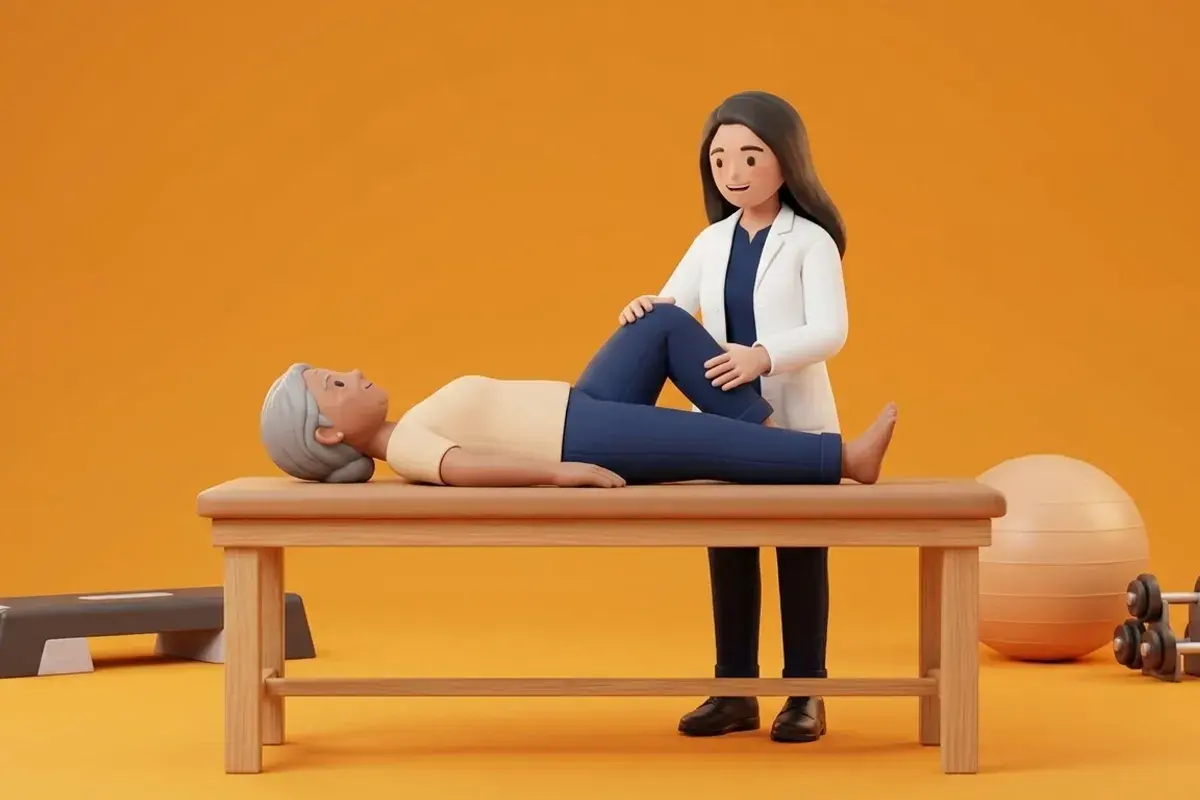
Clinical management of exercise tolerance relies on two non-negotiable tools. First is the stroke test (bulge sign), a validated clinical test to grade knee effusion on a five-point scale (Zero, Trace, 1+, 2+, 3+). The clinician sweeps the medial side of the knee upward toward the suprapatellar pouch two or three times to empty fluid, then sweeps downward along the lateral side; a fluid wave bulging medially reveals an active effusion. Second is the 24-hour symptom response rule: mild pain during exercise (up to 3 or 4 out of 10) is acceptable provided that pain and swelling return to baseline by the following morning. If effusion increases by more than one grade, or morning stiffness exceeds 30 minutes, training loads must be temporarily reduced by 30 to 50 percent, and the PEACE & LOVE protocol (Protection, Elevation, Avoid Anti-inflammatories, Compression, Education, and Load) initiated.
Gentle Supine Passive Heel Slides for Post-Flare Joint Drainage
Objective: Mobilize synovial fluid and stimulate lymphatic drainage without activating inhibited quadriceps muscles during an acute flare
- 1Lie flat on your back on a comfortable bed or exercise mat with your head rested on a pillow.
- 2Loop a long bedsheet, towel, or yoga strap securely around the sole of your affected foot, holding the ends with both hands.
- 3Relax your leg muscles completely, letting your arms do all the physical work of pulling.
- 4Gently pull on the strap with your arms to slide your heel toward your buttock, bending your knee only to a comfortable, pain-free angle.
- 5Slowly lower the foot back down to the straight position over 3 seconds, repeating the passive sliding motion smoothly.
| Effusion Grade | Physical Examination Finding | Joint Status & Muscle Impact | Exercise Prescription Action |
|---|---|---|---|
| Grade Zero (0) | No wave produced on downstroke; joint appears normal and dry | Zero capsular distension; optimal quadriceps motor unit excitability | Green light: Proceed with progressive overload and resistance increases |
| Grade Trace | Small fluid wave appears on medial side with downward lateral stroke | Minimal capsular pressure; mild subclinical synovial reaction | Proceed with current exercise load; do not advance resistance today |
| Grade 1+ | Larger fluid wave returns spontaneously without downward lateral stroke | Moderate capsular distension; 20% to 30% quadriceps inhibition active | Yellow light: Reduce training volume by 30%; avoid deep knee flexion |
| Grade 2+ to 3+ | Fluid wave so large it cannot be swept out; tense suprapatellar fullness | Severe capsular distension; profound quadriceps shutdown and giving way | Red light: Cease resistance; initiate PEACE protocol, cryotherapy, and rest |
An acute, massive joint effusion that develops within 2 to 4 hours following minor movement, accompanied by severe resting throbbing, skin redness, and systemic fever (>38 degrees Celsius), must be evaluated immediately by an orthopaedic surgeon for acute septic arthritis or acute crystalline gout. Immediate sterile joint aspiration for synovial fluid analysis is required.
Frequently Asked Questions
Should I put ice or heat on my knee when it swells up after exercise?
When your knee suddenly swells up with fluid and feels warm after exercise, ice (cold therapy) is the correct choice. Applying an ice pack wrapped in a thin towel for 15 to 20 minutes constricts dilated blood vessels, reduces joint inflammation, and numbs pain. Heat should only be used on stiff, non-swollen muscles before exercise to warm up the joint, never on an actively swollen, hot knee flare.
Do I need a doctor referral to see a knee osteoarthritis physiotherapist on BookPhysio.in?
BookPhysio.in does not require a doctor referral. Under the National Commission for Allied and Healthcare Professions (NCAHP) Act 2021, patients across India enjoy direct access to autonomous registered physiotherapists. Through BookPhysio.in, you connect directly with verified clinicians and pay per session with transparent fees, completely free from advance package commitments, commission markups, or platform subscription charges.
Related condition guides



Related recovery guides & articles
BookPhysio.in Editorial Team
The BookPhysio.in editorial team writes and maintains this content, checking it against reliable clinical sources. Content follows our medical review policy: general information only, and no page claims a clinical review until a named registered physiotherapist has signed it off.
Read our medical review policy