
Monitoring Radiographic Bone Union and Callus Maturity Signs Post Ankle Fracture
In short
Understand radiographic signs of osseous consolidation, cortical bridging callus maturity, and mortise stability following ankle fracture surgery.
In this guide (4 sections)
Evaluating radiographic bone union represents the primary foundation upon which all post-operative weight-bearing progressions, manual therapy mobilizations, and athletic clearance decisions are grounded. Following surgical fixation or conservative immobilization of an ankle fracture, serial radiographic imaging tracks the complex biological transformation of an unstable fracture hematoma into mature, lamellar cortical bone. For the physiotherapist, understanding the precise radiographic markers of osseous consolidation, cortical bridging, and syndesmotic mortise stability is essential. Relying solely on chronological timelines (such as assuming all fractures heal in six weeks) is clinically irresponsible, as bone union rates vary dramatically based on fracture morphology, patient age, vascularity, systemic comorbidities, and fixation rigidity.
Navigating Follow-Up Hospital Visits, Imaging Queues, and Public Transit
In Indian healthcare systems, obtaining serial follow-up radiographs involves practical domestic coordination. Patients must travel from their homes to diagnostic imaging centers or tertiary hospital outpatient departments, often traveling via bumpy auto-rickshaws, crowded cabs, or buses while still managing crutches. Negotiating long hospital corridors, standing in imaging registration queues, and transferring onto cold, elevated X-ray examination tables requires physical endurance and balance. Families often experience anxiety while awaiting radiographic reports, seeking clear explanations of whether the bone has united sufficiently to discard walking aids. Physiotherapists play a vital educational role, helping patients interpret their imaging progress and aligning daily domestic activities with verified biological healing.
Primary Versus Secondary Healing, Cortical Bridging, and Mortise Clear Spaces
The radiographic appearance of bone healing depends fundamentally on the biomechanical stability of the surgical construct. Under absolute stability (achieved with anatomic reduction and rigid compression plating), bone heals via primary (direct) cortical healing without external callus formation; radiographic union is characterized by gradual disappearance of the radiolucent fracture line as osteonal cutting cones cross the contact gap. Under relative stability (typical of bridge plating or comminuted fractures), secondary bone healing occurs via external bridging callus. Serial radiographs (anteroposterior, lateral, and mortise views) are evaluated for three classic criteria: bridging callus crossing at least three of four cortices, continuous trabecular patterns traversing the fracture gap, and complete obliteration of the fracture line. Concurrently, the mortise view is scrutinized: the medial clear space must remain symmetrical and less than four millimeters, confirming syndesmotic integrity.
Radiographic Scoring Systems: The RUST Score, Weight Advancement, and Clinical Checks
Physiotherapists utilize validated objective scoring tools, such as the Radiographic Union Score for Tibia (RUST) adapted for distal lower extremity fractures, to guide functional rehabilitation. The RUST system assigns zero to three points across each cortex based on the presence of bridging callus and fracture line visualization, yielding a score between four (no union) and twelve (complete solid remodeling). When the RUST score reaches seven or eight (bridging callus on at least two cortices without fracture line displacement), partial weight-bearing is initiated. Advancement to full, unrestricted weight-bearing and high-impact activities requires a RUST score of ten or greater, accompanied by clinical union markers: completely painless full weight-bearing, absence of localized bone tenderness upon firm thumb palpation, and lack of resting edema.
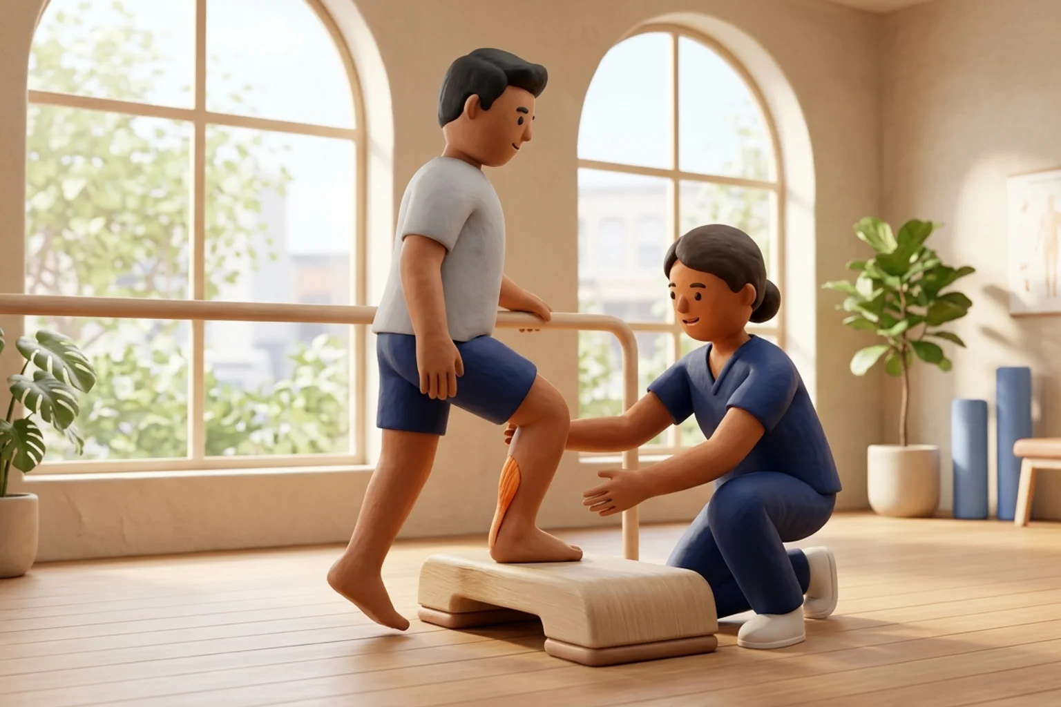
Gentle Axial Loading Weight Transfer Drill with Radiographic Clearance
Objective: Safely stimulate osteoblast bone deposition through controlled, painless physiological Wolff's law loading
- 1Confirm that your orthopaedic surgeon has verified radiographic bridging callus on your latest follow-up X-rays.
- 2Stand upright between two sturdy kitchen counters or parallel bars, resting your hands flat on the surfaces for balance.
- 3Position your feet shoulder-width apart, initially resting seventy percent of your weight on your uninjured leg.
- 4Slowly and gently transfer approximately forty to fifty percent of your body weight onto your recovering foot, holding for five seconds.
- 5Focus on feeling a firm, comfortable pressure across the sole of your foot without experiencing any sharp bone aching.
- 6Transfer weight back to the sound leg, rest for three seconds, and repeat the gentle loading cycle smoothly.
| Radiographic Healing Stage | X-Ray Appearance & Callus Status | Biomechanical Bone Strength | Permitted Functional Milestone |
|---|---|---|---|
| Early Healing (Weeks 2-6) | Sharp fracture lines visible; early faint periosteal haze | Minimal; relies entirely on metallic plates and screws | Strict Non-Weight-Bearing; gentle active ROM in boot |
| Progressive Union (Weeks 6-10) | Bridging callus visible across 2-3 cortices; lines fading | Moderate; bone accepts progressive axial compression | Partial weight-bearing (25-75%); crutch weaning; balance drills |
| Consolidated Union (Weeks 10-16+) | Bridging across 4 cortices; dense lamellar remodeling | Near-normal physiological strength restored | Full independent walking; agility drills; low-impact sports |
Carefully inspect follow-up radiographs for a thin, continuous radiolucent halo surrounding metallic screw threads or under plate surfaces. A radiolucent line greater than one millimeter wide indicates micro-motion, hardware loosening, or peri-implant osteolysis. If accompanied by localized pain during weight-bearing, axial loading must be halted immediately and referred for urgent orthopaedic review.
Frequently Asked Questions
Why does my X-ray still show a faint fracture line even though my ankle feels completely fine?
Bone remodeling is a slow biological process that takes up to one to two years to completely erase a fracture line on an X-ray. As long as your X-ray shows solid bridging bone (callus) connecting the fragments and you have no pain when walking, your bone is structurally strong enough to support your full body weight.
Do I need a doctor referral to rehabilitate an ankle fracture on BookPhysio.in?
BookPhysio.in does not require a doctor referral. Under the National Commission for Allied and Healthcare Professions (NCAHP) Act 2021, patients across India enjoy direct access to autonomous registered physiotherapists. Through BookPhysio.in, you connect directly with verified clinicians and pay per session with transparent fees, completely free from advance package commitments, commission markups, or platform subscription charges.
Related condition guides



Related recovery guides & articles
BookPhysio.in Editorial Team
The BookPhysio.in editorial team writes and maintains this content, checking it against reliable clinical sources. Content follows our medical review policy: general information only, and no page claims a clinical review until a named registered physiotherapist has signed it off.
Read our medical review policy