
Patellar Mobilization in Knee Osteoarthritis: Superior, Inferior, and Transverse Glides
In short
Master manual patellar mobilization techniques to restore patellofemoral arthrokinematics, free retinacular adhesions, and eliminate joint stiffness in knee OA.
In this guide (4 sections)
In the comprehensive physiotherapy management of knee osteoarthritis, restoring normal accessory gliding of the patella is a fundamental manual therapy prerequisite for restoring active physiological joint motion. The patellofemoral joint is a modified planar articulation where the articular surface of the patella must glide smoothly through the femoral trochlear groove during knee flexion and extension. Specifically, the patella glides inferiorly (distally) during knee flexion and superiorly (proximally) during knee extension, accompanied by subtle medial and lateral tilts. In knees affected by osteoarthritis, recurrent joint effusions, quadriceps hypertonicity, and chronic disuse lead to severe hypomobility of the patella. The peripatellar retinacula and joint capsule develop dense fibrous adhesions that effectively tether the patella, locking it in an abnormal lateral tilt or superior displacement. Manual patellar mobilization using calibrated superior, inferior, and transverse glides restores joint arthrokinematics, reduces retinacular tension, and alleviates retro-patellar grinding pain.
Indian Household Floor Transfers: Sitting on Mats and Reaching Overhead
In Indian daily routines, patellar hypomobility severely impairs practical domestic function. When an individual attempts to sit down on a prayer mat or rise from a low diwan, the knee must bend past 90 degrees; if the patella is tethered and cannot glide freely in an inferior direction, extreme compressive shear is generated across the patellofemoral joint, provoking sharp anterior knee pain and crepitus. Similarly, reaching up to high kitchen storage shelves while standing requires full knee extension; if superior patellar glide is restricted, the knee cannot achieve full extension, leading to compensations in the lumbar spine. Teaching patients gentle self-mobilization techniques that can be performed at home in bed or on a comfortable sofa empowers them to maintain daily joint freedom.
Arthrokinematic Coupling, Retinacular Contracture, and Trochlear Entrapment
The biomechanical necessity of patellar mobility is governed by arthrokinematic rules. During full knee flexion to 135 degrees, the patella must travel inferiorly by roughly 5 to 7 centimeters to seat congruently in the intercondylar notch. If the superior retinaculum or rectus femoris is contracted, inferior glide is blocked, causing the patella to act as a rigid mechanical wedge that prematurely impacts the anterior femoral condyle. Conversely, during terminal knee extension, the patella must glide freely superiorly; if the patellar tendon is shortened or the infrapatellar fat pad is inflamed and fibrotic, superior glide is restricted, preventing the vastus medialis oblique from achieving full terminal extension lockout. Restoring transverse medial glide is equally vital: because the lateral retinaculum is typically thickened in osteoarthritis, restoring medial translation decompresses the chronically overloaded lateral patellar facet.
Manual Mobilization Grades (Maitland I to IV), Directional Glides, and Patient Relaxation
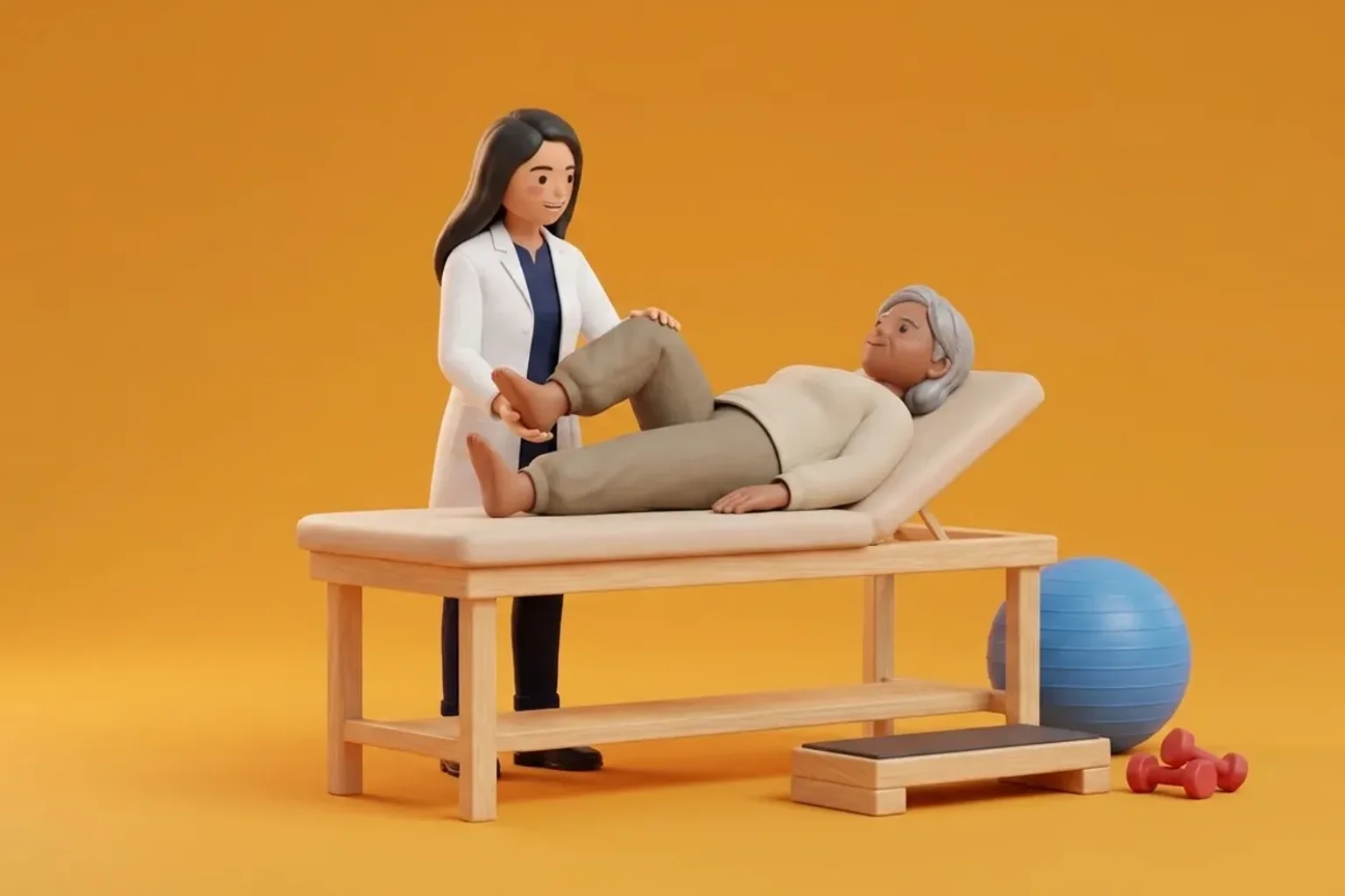
Clinical execution of patellar mobilization requires complete patient relaxation with the quadriceps fully disengaged. The patient lies supine with a small towel roll placed beneath the popliteal fossa to keep the knee in roughly 15 to 20 degrees of resting flexion. Clinicians utilize Maitland passive mobilization grades: gentle low-amplitude oscillations (Grades I and II) are applied for neurophysiological pain modulation and synovial lubrication, while sustained, large-amplitude stretches (Grades III and IV) are applied at the end-range of capsular restriction to induce mechanical tissue lengthening. Mobilizations are applied systematically in superior, inferior, and medial directions, avoiding excessive lateral translation which could aggravate lateral patellar instability.
Seated Self-Administered Medial Patellar Glide Mobilization
Objective: Mobilize the patella medially to stretch contracted lateral retinacular tissues and decompress the lateral patellar facet
- 1Sit comfortably on a firm bed or sofa with your affected leg extended completely straight out in front of you, supported by the mattress.
- 2Completely relax your thigh muscles, verifying that your kneecap is loose and can be easily moved with your fingers.
- 3Place the base of your thumbs along the outer (lateral) border of your kneecap.
- 4Gently and smoothly push your kneecap inward toward the midline of your body until you feel a firm, comfortable resistance stretch.
- 5Hold the medial stretch for 5 seconds, release slowly, and repeat in a gentle rhythmic oscillating manner for 2 minutes.
| Glide Direction | Primary Arthrokinematic Role | Targeted Anatomical Restriction | Clinical Indication in Knee OA |
|---|---|---|---|
| Inferior (Distal) Glide | Essential for full knee flexion; guides patella into trochlear groove | Tight quadriceps tendon, rectus femoris, and superior capsule | Inability to bend knee past 90 degrees; severe pain during squatting |
| Superior (Proximal) Glide | Essential for full terminal knee extension and screw-home lockout | Tight patellar tendon, fibrotic infrapatellar fat pad, inferior capsule | Persistent extensor lag; inability to straighten leg fully while walking |
| Medial Transverse Glide | Decompresses lateral patellofemoral facet; stretches lateral retinaculum | Dense, contracted lateral patellar retinaculum and iliotibial band fibers | Severe lateral facet osteoarthritis, patellar tilt, and anterior knee pain |
| Lateral Transverse Glide | Maintains medial retinacular compliance and capsular mobility | Applied cautiously; avoids excessive stretching of medial stabilizers | Used selectively for generalized patellar stiffness after prolonged casting |
Patellar mobilizations must be discontinued immediately if the patient reports severe sharp pain or apprehension suggesting acute patellofemoral subluxation. Furthermore, manual mobilizations are strictly contraindicated in the presence of acute patellar fracture, severe prepatellar bursitis, active cellulitis, or acute deep vein thrombosis.
Frequently Asked Questions
Why does my physiotherapist move my kneecap around with their fingers before having me exercise?
Your kneecap needs to slide smoothly up and down along the thigh bone groove for your knee to bend and straighten without pain. In osteoarthritis, the fibrous bands around the kneecap become tight and stiff, trapping the kneecap in place like a rusty hinge. By manually gliding your kneecap before you exercise, your physiotherapist frees up these tight tissues and circulates joint fluid, allowing you to exercise with much less pain.
Do I need a doctor referral to see a knee osteoarthritis physiotherapist on BookPhysio.in?
BookPhysio.in does not require a doctor referral. Under the National Commission for Allied and Healthcare Professions (NCAHP) Act 2021, patients across India enjoy direct access to autonomous registered physiotherapists. Through BookPhysio.in, you connect directly with verified clinicians and pay per session with transparent fees, completely free from advance package commitments, commission markups, or platform subscription charges.
Related condition guides

Related recovery guides & articles
BookPhysio.in Editorial Team
The BookPhysio.in editorial team writes and maintains this content, checking it against reliable clinical sources. Content follows our medical review policy: general information only, and no page claims a clinical review until a named registered physiotherapist has signed it off.
Read our medical review policy